For decades, the central promise of regenerative medicine—the ability to grow replacement human organs, muscle flaps, and vascular networks in the laboratory to cure heart failure, severe trauma, or paralysis—was hindered by a formidable physical barrier: the diffusion limit of oxygen. In all complex multicellular living mammalian tissues, metabolically active cells situated more than 200 micrometers (0.2 mm) from an active capillary bed rapidly suffocate and undergo ischemic necrosis. Without an integrated, functional circulatory network of microscopic blood vessels, engineered 3D biological tissues were doomed to remain paper-thin cellular sheets.
Israeli biomedical engineer and stem cell biophysicist Professor Shulamit Levenberg shattered this biological bottleneck. Conducting seminal research at the Massachusetts Institute of Technology (MIT) alongside Institute Professor Robert Langer and continuing at the Technion – Israel Institute of Technology in Haifa, Levenberg pioneered the engineering of pre-vascularized 3D tissue constructs. By co-culturing endothelial cells, supporting pericytes/fibroblasts, and target tissue-specific stem cells on biodegradable polymeric scaffolds, her laboratory created self-assembling capillary networks that rapidly anastomose (connect) with host blood vessels upon transplantation.
Beyond regenerating human heart muscle, cranial bone, and severed spinal cords, Levenberg applied her 3D vascularization and scaffold technology to revolutionize global food systems. As co-founder and Chief Scientific Advisor of Aleph Farms, she developed the world’s first slaughter-free, 3D-structured cultivated beef steaks, establishing Israel as the undisputed epicenter of sustainable cellular agriculture. Recognized by Scientific American as one of the 50 world leaders in science and technology, Professor Levenberg stands at the forefront of modern bio-convergence. This definitive study explores her scientific journey, bioengineering breakthroughs, regenerative therapies, and visionary contributions to sustainable biotechnology.
1. Academic Journey: From Weizmann Institute Biophysics to MIT
Shulamit Levenberg was born and raised in Israel during an era of rapid technological and scientific expansion. Exhibiting exceptional intellectual acuity in mathematics and life sciences from an early age, she enrolled at the Hebrew University of Jerusalem, graduating with a Bachelor of Science (B.Sc.) in Biology with distinction in 1993.
She then joined the world-renowned Weizmann Institute of Science in Rehovot to pursue direct doctoral studies in cell biology under Professor Benjamin Geiger. Her Ph.D. research (completed in 1999) unraveled the molecular mechanisms of cell adhesion, focal contacts, and cell-matrix signaling mediated by cadherins and integrins—biochemical knowledge that would later prove crucial for engineering synthetic extracellular matrix environments.
Postdoctoral Breakthroughs with Robert Langer at MIT
Between 1999 and 2004, Dr. Levenberg conducted groundbreaking postdoctoral research at the Massachusetts Institute of Technology (MIT) in the laboratory of Institute Professor Robert Langer, the world’s most cited engineer and a founding father of modern biomaterials and drug delivery systems.
Working in collaboration with stem cell researchers at Harvard University and MIT, Levenberg investigated how human embryonic stem cells (hESCs) could be guided to differentiate into complex three-dimensional tissues. She discovered that by seeding stem cells into porous, highly flexible biodegradable polymer scaffolds composed of poly(L-lactic acid) (PLLA) and poly(lactic-co-glycolic acid) (PLGA), mechanical forces and matrix geometry prompted cells to spontaneously assemble into multi-cellular architectures resembling developing embryonic tissues.
| Academic Stage | Institution | Year | Research Focus / Landmark Achievement |
|---|---|---|---|
| B.Sc. Biology (Honors) | Hebrew University of Jerusalem | 1993 | Cellular physiology, biochemistry, genetics. |
| Ph.D. Cell Biology | Weizmann Institute of Science | 1999 | Molecular mechanisms of cell-cell adhesion and cadherin signaling under Benjamin Geiger. |
| Postdoctoral Fellow | Massachusetts Institute of Technology (MIT) | 1999–2004 | Stem cell differentiation and vascularized 3D tissue engineering under Robert Langer. |
| Faculty Member & Dean | Technion – Faculty of Biomedical Engineering | 2004–Present | Director of Stem Cell and Tissue Engineering Laboratory; Dean of Biomedical Engineering (2017–2021). |
| Co-Founder & Chief Scientist | Aleph Farms Ltd. | 2017–Present | Pioneered world’s first 3D-structured cultivated beef steaks using non-GMO soy scaffolds. |
2. The Vascularization Dilemma: Overcoming the 200-Micron Diffusion Barrier
Every mammalian tissue requires a continuous supply of oxygen, glucose, and essential nutrients, alongside the rapid clearance of metabolic waste products like lactate and carbon dioxide. In biological systems, passive diffusion is effective only over distances of **100 to 200 micrometers** (roughly the width of two human hairs).
Prior to Levenberg’s inventions, when tissue engineers attempted to culture thicker 3D muscle or liver grafts (1–5 mm thick), cells in the interior suffered rapid hypoxia, nutrient starvation, and central necrosis within 24–48 hours. Furthermore, even if thick tissues survived briefly in vitro within oxygenated bioreactors, upon surgical implantation in vivo, host blood vessels took weeks to slowly sprout into the graft (angiogenesis), by which time the engineered graft had largely died.
The Co-Culture Triad Strategy
In a landmark 2005 publication in Nature Biotechnology, Levenberg revealed her revolutionary solution: pre-vascularization prior to transplantation. Instead of seeding a scaffold with a single cell type, Levenberg designed a tri-culture cellular system:
- Human Umbilical Vein Endothelial Cells (HUVECs): The building blocks of blood vessel linings.
- Embryonic Fibroblasts / Pericytes: Support cells that secrete extracellular matrix and stabilize vessel walls with smooth muscle-like mural coverage.
- Skeletal Myoblasts or Cardiomyocytes: The functional parenchymal muscle cells.
When seeded together onto porous PLLA/PLGA biodegradable scaffolds, the endothelial cells spontaneously organized into branching, tubular lumen structures lined with tight junctions, surrounded by pericytes and mature muscle fibers. When these pre-vascularized tissue flaps were transplanted into surgical muscle defects in rodents, the engineered capillaries underwent rapid anastomosis—physically fusing with the host circulatory system within **48 to 72 hours**, establishing full blood perfusion and 100% graft survival.
3. The Biomechanical Engineering of Biomaterial Scaffolds
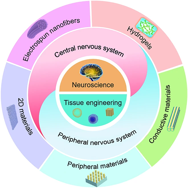
Engineering functional 3D biological tissues requires far more than mixing cells in a Petri dish; it demands the design of highly customized extracellular matrix (ECM) scaffolds that provide structural support, biochemical signaling, and precise mechanical cues.
1. Polymer Chemistry and Degradation Kinetics
Professor Levenberg’s laboratory engineered synthetic and natural polymeric matrices tailored to specific tissue microenvironments:
- Poly(L-lactic acid) (PLLA) & Poly(lactic-co-glycolic acid) (PLGA): Synthetic aliphatic polyesters whose hydrolytic ester cleavage rates can be fine-tuned from weeks to months by altering the lactic-to-glycolic acid copolymer ratio (e.g., PLGA 50:50 versus PLGA 85:15). Degradation products (lactic acid and glycolic acid) are naturally metabolized via the Krebs cycle without cytotoxic accumulation.
- Methacrylated Gelatin (GelMA) & Fibrin Hydrogels: Photopolymerizable hydrogels crosslinked via ultraviolet or blue light (405 nm) with LAP photoinitiators, replicating the soft, hydrated viscoelastic environment of native human soft tissues (1–10 kPa stiffness).
- Decellularized Extracellular Matrix (dECM): Natural organ matrices stripped of all cellular antigens through detergent washing (Triton X-100, sodium deoxycholate), preserving native tissue-specific architecture, glycosaminoglycans, and growth factor reservoirs.
2. Scaffold Fabrication and Pore Architecture
To enable cellular migration and nutrient exchange, scaffolds must exhibit interconnecting porosities exceeding 85% with specific pore diameters:
- Electrospinning: Utilizes high-voltage electrostatic fields (15–25 kV) to draw polymer solutions into micro- and nanoscale fibrous meshes mimicking the fibrous collagen architecture of native muscle and neural tracts.
- Particulate Salt Leaching: Incorporates calibrated sodium chloride crystals into polymer solutions, which are subsequently dissolved in water to generate highly interconnected spherical pore geometries (150–300 μm).
- Extrusion and Digital Light Processing (DLP) 3D Bioprinting: Layer-by-layer deposition of cell-laden bioinks with micrometer-scale spatial resolution.
| 3D Bioprinting Modality | Mechanism / Physical Principle | Resolution & Speed | Primary Tissue Application |
|---|---|---|---|
| Pneumatic / Piston Extrusion | Continuous mechanical deposition of shear-thinning hydrogels. | 100–500 μm | Moderate speed | Thick vascularized muscle constructs, cultivated meat scaffolds, bone graft implants. |
| Digital Light Processing (DLP) | Photopolymerization of liquid bioinks using dynamic micromirror projection. | 10–50 μm | Ultra-fast | High-precision microfluidic capillary channels, complex vascular branching trees, neural guides. |
| Laser-Assisted Bioprinting (LAB) | Pulsed laser-induced forward transfer of microscopic cell droplets. | 10–100 μm | Moderate speed | High cell-viability skin patterning, delicate corneal cellular layers. |
| Coaxial Microfluidic Extrusion | Concentric nozzle extrusion forming hollow tubular conduits on-the-fly. | 50–200 μm | Fast | Perfusible micro-vessel channels, renal proximal tubules, gut-on-a-chip conduits. |
4. Microvascular Fluid Dynamics: Inosculation vs. Angiogenesis
Professor Levenberg’s biophysical studies clarified the exact cellular mechanisms distinguishing standard host angiogenesis from engineered inosculation:
The Molecular Mechanics of Capillary Fusion
- Slow Host Sprouting (Angiogenesis): When an avascular graft is transplanted, host endothelial cells must slowly degrade host basement membrane, proliferate, and migrate into the graft at a sluggish pace of ~5 μm/hour. In thick grafts, interior cells die long before host vessels reach them.
- Engineered Pre-Vascular Inosculation: In Levenberg’s pre-vascularized constructs, fully formed lumenized capillary networks already exist throughout the graft. Upon transplantation, the open capillary ends of the graft physically dock and fuse (inosculate) with sprouting host capillaries across the entire graft boundary within 24 to 48 hours.
- Tip-Cell vs. Stalk-Cell Dynamics: Endothelial morphogenesis in the scaffold is regulated by Notch-Delta (Dll4-Notch1) lateral inhibition signaling, establishing leading exploratory tip cells guided by localized VEGF-A gradients, followed by proliferating stalk cells that assemble patent hollow lumens.
5. Regenerative Medicine Frontiers: Spinal Cord Repair and Cardiac Flaps
At the Technion, Professor Levenberg expanded her pre-vascularized tissue engineering platform to tackle some of the most devastating conditions in human clinical medicine:
1. Complete Spinal Cord Injury Repair
Traumatic spinal cord severance causes permanent paralysis because adult central nervous system neurons cannot regenerate across glial scar tissue and fluid-filled cyst cavities. In a series of groundbreaking studies published in Frontiers in Neuroscience and Biomaterials, Levenberg’s team engineered functional 3D neural tissue implants:
- Porous biodegradable scaffolds were seeded with human Dental Pulp Stem Cells (hDPSCs) and neural progenitor cells, which secreted high levels of neurotrophic factors (BDNF, GDNF, and VEGF).
- When implanted into adult rats with complete spinal cord transection, the engineered neural scaffolds bridged the gap, guided directional axonal regeneration across the lesion, promoted remyelination by oligodendrocytes, and induced significant functional motor recovery (hindlimb walking gait).
2. Pre-Vascularized Cardiac Muscle Patches
Following myocardial infarction (heart attack), millions of cardiac muscle cells die and are replaced by non-contractile fibrotic scar tissue, leading to congestive heart failure. Levenberg engineered thick, electromechanically coupled cardiac muscle patches incorporating human induced pluripotent stem cell-derived cardiomyocytes (hiPSC-CMs) and endothelial networks. Upon transplantation onto infarcted rodent hearts, these patches integrated electrically with host myocardium, improved left ventricular ejection fraction, and prevented adverse ventricular remodeling.
3. Osteogenic Flaps for Craniofacial Bone Reconstruction
Treating massive composite craniofacial bone defects resulting from blast trauma or oncological resection requires both vascularized bone and soft tissue coverage. Levenberg developed engineered osteogenic-endothelial constructs that synthesize mineralized hydroxyapatite matrix in vitro and rapidly vascularize upon implantation, providing a clinically translatable blueprint for reconstructive plastic surgery.
| Clinical / Medical Application | Scaffold Material | Cellular Components | Therapeutic Mechanism & Outcome |
|---|---|---|---|
| Spinal Cord Neuroregeneration | Electrospun PLLA/PLGA aligned fibers | Dental Pulp Stem Cells (hDPSCs) + Neural Progenitors | Bridges severed spinal cord, secretes neurotrophic factors, restores motor locomotion in animal models. |
| Myocardial Infarction Patch | Decellularized extracellular matrix / Porous PLGA | hiPSC Cardiomyocytes + Endothelial Cells + Pericytes | Electromechanically synchronizes with native heart tissue; improves cardiac output and reduces scar size. |
| Composite Skeletal Muscle Flap | Porous sponge PLLA / Fibrin gel hydrogel | Human Skeletal Myoblasts + HUVECs + Fibroblasts | Rapid host vascular anastomosis within 48h; repairs massive volumetric muscle loss (VML). |
| Craniofacial Bone Regeneration | Hydroxyapatite-coated polycaprolactone (PCL) | Mesenchymal Stem Cells (MSCs) + Endothelial Networks | Induces robust vascularized osteogenesis, repairing critical-sized segmental bone voids. |
6. Cellular Agriculture & Aleph Farms: Cultivating the Future of Food
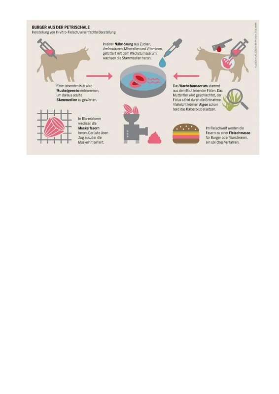
Traditional industrial cattle farming is one of the world’s leading drivers of greenhouse gas emissions, deforestation, freshwater depletion, and antibiotic resistance. In 2017, Professor Levenberg realized that the identical 3D tissue engineering principles she had pioneered for human medicine could be harnessed to produce slaughter-free, authentic animal meat.
Founding Aleph Farms
In partnership with food-tech incubator The Kitchen Hub (Strauss Group) and entrepreneur Didier Toubia, Levenberg co-founded Aleph Farms in Rehovot, Israel, serving as Chief Scientific Advisor. While early cultivated meat startups produced unstructured cell purees suitable only for processed burgers or nuggets, Aleph Farms aimed for the ultimate culinary holy grail: thick, 3D-structured beef steaks containing genuine muscle fibers, fat cells, and connective tissue.
The Textured Soy Protein Scaffold Innovation
In 2018, Aleph Farms made global history by unveiling the world’s first slaughter-free cultivated thin-cut beef steak. In 2021, Levenberg’s laboratory published the definitive bioprocess blueprint in Nature Food:
- Non-GMO Textured Soy Protein (TSP): Instead of synthetic medical polymers, Levenberg utilized edible, porous textured soy protein as a biocompatible scaffold. The microscopic pores provide ideal mechanical stiffness and topography for bovine cells to adhere, proliferate, and align.
- Co-Culture of Four Bovine Cell Types: Non-immortalized, non-genetically modified bovine satellite cells (muscle), pre-adipocytes (fat), fibroblasts (collagen matrix), and endothelial-like cells were co-cultured inside the soy scaffold.
- Serum-Free Culture Medium: Eliminated all fetal bovine serum (FBS), replacing it with animal-free, food-grade amino acids, lipids, and plant-derived growth factors.
- Texture, Taste, and Culinary Performance: The resulting cultivated steak replicated the marbling, mouthfeel, bite resistance, and sizzle of conventional pasture-raised beef when cooked, while requiring up to 95% less land, 78% less water, and generating 92% fewer carbon emissions.
10. Space Biotechnology: Cultivating Meat on the International Space Station
In September 2019, Aleph Farms and Professor Levenberg achieved another historic world first: cultivating meat in space. Conducting a joint experiment aboard the International Space Station (ISS) in collaboration with 3D Bioprinting Solutions, bovine cells were bioprinted into muscle tissue under microgravity conditions 400 kilometers above Earth.
In 2022, Aleph Farms expanded its space research during the Rakia Mission aboard the ISS with Israeli astronaut Eytan Stibbe, analyzing how microgravity influences bovine stem cell proliferation and tissue assembly. The technology offers a viable closed-loop nutritional solution for long-duration deep space exploration missions to the Moon and Mars.
| Environmental Metric | Conventional Cattle Farming | Aleph Farms Cultivated Beef | Environmental / Societal Benefit |
|---|---|---|---|
| Land Usage | ~250–350 m² per kg beef | ~15 m² per kg beef | 95% reduction in agricultural land use; halts Amazon rainforest deforestation. |
| Freshwater Consumption | 15,400 liters per kg beef | ~3,300 liters per kg beef | 78% reduction in freshwater depletion; climate-resilient food production. |
| Greenhouse Gas Emissions | ~60 kg CO₂-eq per kg beef (enteric methane) | ~5 kg CO₂-eq (renewable energy) | Up to 92% reduction in greenhouse gas emissions; zero enteric methane production. |
| Antibiotic Utilization | Massive preventive herd administration | 100% Antibiotic-Free (Closed Bioreactors) | Eliminates agricultural antimicrobial resistance risk and zoonotic pathogen outbreaks. |
7. Bioprocess Engineering for Scalable Cellular Agriculture
Transitioning from cultivating milligram-scale tissue samples in research Petri dishes to industrial-scale production of metric tons of cultivated beef requires overcoming formidable biochemical and bioprocess engineering challenges. As Chief Scientific Advisor of Aleph Farms, Professor Levenberg guided the development of proprietary bioprocess technologies:
1. Bioreactor Architectures and Hydrodynamic Shear Stress
Mammalian cells lack the rigid peptidoglycan cell walls of bacteria or yeast, making them vulnerable to lethal hydrodynamic shear stresses caused by mechanical impellers. Levenberg’s team optimized specialized bioreactor environments:
- Perfusion Hollow-Fiber Bioreactors: Employs bundles of semi-permeable capillary fibers through which nutrient-rich, oxygenated medium flows continuously, mimicking physiological capillary beds and allowing high-density cell packing without turbulence.
- Low-Shear Stirred-Tank Bioreactors: Utilizes marine-type impellers and computational fluid dynamics (CFD) modeling to maintain homogeneous mixing and bubble-free micro-sparging of dissolved oxygen while keeping shear stresses below the threshold of myoblast membrane rupture.
- Edible Microcarrier Suspension: For cellular expansion phases, non-immortalized bovine cells are cultured on edible, biodegradable microcarriers suspended in gentle fluid motion, maximizing surface-area-to-volume ratios.
2. The Serum-Free Media Revolution
Conventional biomedical cell culture relied heavily on Fetal Bovine Serum (FBS)—a costly, non-standardized animal product that contradicted the ethical and economic goals of cultivated meat. Aleph Farms achieved 100% serum-free production by engineering a defined food-grade medium consisting of:
- Recombinant Growth Factors: Precision-fermented basic fibroblast growth factor (bFGF / FGF-2) and transforming growth factor-beta (TGF-beta) produced in yeast or microalgae.
- Plant and Algal Hydrolysates: Sustainable peptide sources derived from soy, pea, and spirulina providing essential amino acids and trace minerals.
- Metabolic Waste Clearance: Continuous membrane filtration systems that extract toxic ammonia and lactate byproducts while recycling expensive vitamins and fatty acids, slashing media costs by over 90%.
| Production Characteristic | Conventional Livestock Beef | Plant-Based Meat Substitutes | Aleph Farms Cultivated Beef |
|---|---|---|---|
| Cellular Composition | Intact animal muscle, adipose, and connective tissues. | Processed soy, pea, or wheat plant proteins with binders/oils. | Authentic bovine myoblasts, pre-adipocytes, fibroblasts, and collagen. |
| 3D Tissue Architecture | Natural skeletal muscle bundle architecture. | Extruded paste or homogenized particulate texture. | Structured 3D myofibers aligned within textured plant protein scaffold. |
| Production Timeline | 18–30 months per cattle harvest. | Agricultural crop growing seasons (months). | 3 to 4 weeks inside sterile bioreactor cultivators. |
| Pathogen & Contamination Risk | High risk of *Salmonella*, *E. coli* O157:H7, campylobacter, and bovine spongiform encephalopathy. | Moderate risk of agricultural mycotoxins and pesticide residues. | Sterile pharmaceutical-grade cleanroom environment; zero fecal or zoonotic contamination. |
8. Advanced Mechanobiology: How Physical Forces Dictate Stem Cell Destiny
A core pillar of Professor Levenberg’s scientific legacy is her pioneering work in cellular mechanobiology—unraveling how stem cells physically “feel” their microenvironment and convert mechanical forces into genomic expression programs:
The Stiffness-Lineage Mapping
Levenberg demonstrated that stem cell differentiation is governed not merely by chemical growth factors, but by the physical elastic modulus (Young’s modulus, $E$) of the surrounding scaffold matrix:
- Ultra-Soft Scaffolds ($E sim 0.1 ext{–}1 ext{ kPa}$): Mimicking native brain and spinal tissue; promotes differentiation of neural progenitor cells into neurons and glial cells.
- Intermediate Stiffness Scaffolds ($E sim 8 ext{–}15 ext{ kPa}$): Mimicking mammalian skeletal muscle; activates myogenic transcription factors (MyoD, Myogenin), driving myoblasts to fuse into multi-nucleated, contractile myotubes.
- Rigid Scaffolds ($E sim 30 ext{–}100 ext{ kPa}$): Mimicking collagenous pre-bone osteoid; triggers Runx2 and osteocalcin expression, driving robust osteogenic differentiation and hydroxyapatite mineralization.
The YAP/TAZ Mechanotransduction Cascade
At the molecular level, mechanical tension transmitted through focal adhesions and integrins activates RhoA/ROCK signaling, reorganizing the actin cytoskeleton. This prevents phosphorylation of the transcriptional co-activators YAP (Yes-associated protein) and TAZ, permitting their translocation into the nucleus where they bind TEAD transcription factors to execute tissue-specific differentiation and proliferation programs.
9. The Technion 3D Bioprinting Center and the Israeli Bio-Convergence Hub
Under Professor Levenberg’s vision, the Technion established the Technion Center for 3D Bioprinting, equipped with state-of-the-art multi-material bioprinters, laser sintering systems, micro-computed tomography (micro-CT), and bioreactor development suites.
The center serves as the core anchor of Israel’s national Bio-Convergence Strategy, orchestrated by the Israel Innovation Authority. By bridging engineering, biology, nanotechnology, and advanced computer algorithms, Levenberg’s academic hub has incubated a vibrant cluster of medtech and food-tech ventures, training a new generation of interdisciplinary biomedical scientists driving Israel’s innovation economy.
11. Global Regulatory Milestones and Kosher / Halal Certification
The transition of cultivated meat from academic curiosity to commercial reality required navigating unprecedented regulatory, safety, and religious frameworks:
1. World-First Regulatory Approval for Cultivated Beef
In January 2024, the Israeli Ministry of Health granted historic regulatory approval to Aleph Farms for its cultivated beef steak (marketed under the brand name Aleph Cuts). This landmark decision made Israel the first country in the world to authorize the sale of cultivated beef, and only the third country globally (after Singapore and the United States) to approve any form of cultivated meat for public consumption.
2. Halachic and Islamic Jurisprudence Breakthroughs
Cultivated meat raised profound theological questions for religious dietary laws:
- Historic Kosher Ruling: In January 2023, the Ashkenazi Chief Rabbi of Israel, Rabbi David Lau, issued a historic halachic ruling declaring that cultivated beef produced by Aleph Farms using stem cells harvested from a fertilized egg is Kosher (Pareve/Fleishig), provided strict supervision protocols are maintained. This opened immense global kosher consumer markets.
- Halal Compatibility: Leading Islamic scholars and halal certification authorities in the United Arab Emirates, Saudi Arabia, and Southeast Asia subsequently confirmed that cultivated meat is Halal-compliant when derived from halal animals and cultured in strictly porcine-free, alcohol-free, and clean nutrient media.
12. Future Frontiers: 4D Bioprinting, Smart Biomaterials, and Organ-on-a-Chip
Looking toward the future, Professor Levenberg’s laboratory at the Technion is spearheading the next revolution in biomaterials science:
1. 4D Bioprinting with Stimuli-Responsive Shape-Memory Polymers
Standard 3D bioprinting produces static structures. In contrast, 4D Bioprinting incorporates stimuli-responsive hydrogels that change their physical shape, porosity, or mechanical stiffness over time in response to physiological triggers (such as temperature, pH shifts, enzyme activity, or electrical fields). This enables minimally invasive surgical delivery: collapsed, compressed cellular scaffolds can be injected through micro-catheters and automatically expand into full 3D muscle or vascular grafts inside the body upon reaching 37°C body temperature.
2. Human Microphysiological Organ-on-a-Chip Platforms
By integrating microfluidics with pre-vascularized human tissues, Levenberg’s team is building advanced Organ-on-a-Chip devices (including muscle-on-a-chip, blood-brain-barrier-on-a-chip, and tumor-microenvironment-on-a-chip). These micro-devices allow pharmaceutical companies to evaluate drug efficacy and toxicity directly on human cellular models, drastically accelerating preclinical drug development while phasing out animal testing.
13. Academic Leadership, Technion Dean, and International Honors
Between 2017 and 2021, Professor Shulamit Levenberg served as Dean of the Faculty of Biomedical Engineering at the Technion. Under her leadership, the faculty established the Technion Center for 3D Bioprinting, expanded clinical translation partnerships with Rambam Health Care Campus, and launched pioneering multidisciplinary bio-convergence programs integrating biology, computing, materials science, and nanotechnology.
Major Awards and Distinctions
- Scientific American 50 (2006): Selected as one of the 50 world leaders in science and technology for pioneering 3D tissue vascularization.
- Krill Prize for Excellence in Scientific Research (2008): Awarded by the Wolf Foundation for outstanding achievements in stem cell bioengineering.
- Rappaport Prize for Biomedical Research (2018): Honored for groundbreaking translational contributions to spinal cord regeneration and vascularized tissue engineering.
- Bruno Memorial Award (2018): Granted by the Israel Institute for Advanced Studies to leading Israeli scholars.
- President of the Israel Stem Cell Society: Led national stem cell research standards, international collaborations, and bioethical guidelines.
14. Mentorship, Diversity in STEM, and Entrepreneurial Leadership
As one of Israel’s most prominent female scientific pioneers and tech founders, Professor Shulamit Levenberg is a passionate advocate for empowering women in deep-tech entrepreneurship, engineering, and academia:
Fostering the Next Generation of Bioengineers
Throughout her tenure as Dean of Biomedical Engineering at the Technion, Levenberg actively championed programs to recruit, mentor, and promote female undergraduate and graduate students in engineering disciplines. Her laboratory has trained dozens of Ph.D. candidates and postdoctoral fellows who now hold prestigious faculty positions in leading universities across Israel, North America, and Europe, or lead research and development divisions in cutting-edge biotechnology multinationals.
Bridging the Academic-Commercial Chasm
Levenberg frequently lectures on the vital importance of technology transfer and scientific entrepreneurship. She emphasizes that basic scientific discovery and commercial translation are deeply complementary pursuits: deep-tech entrepreneurship transforms laboratory insights into real-world solutions that save human lives, regenerate failing organs, and protect the global environment for future generations.
15. Frequently Asked Questions (FAQ)
What is Professor Shulamit Levenberg famous for?
Professor Shulamit Levenberg is internationally renowned for solving the vascularization bottleneck in tissue engineering (creating pre-vascularized 3D biological tissues), pioneering spinal cord repair using stem cell scaffolds, and co-founding Aleph Farms to produce the world’s first cultivated beef steaks.
Why was tissue vascularization such a major scientific breakthrough?
Because oxygen and nutrients diffuse only up to 200 micrometers, thicker engineered tissues previously died of hypoxia. Levenberg created self-assembling capillary networks in vitro that connect with host blood vessels within days of transplantation, enabling thick 3D tissue survival.
How does Aleph Farms cultivate 3D beef steaks without slaughter?
Aleph Farms co-cultures authentic, non-genetically modified, non-immortalized bovine muscle satellite cells, fat, and supporting cells inside edible, textured soy protein scaffolds within clean bioreactors, utilizing animal-free nutrients to grow structured steaks that replicate the texture and taste of beef.
What progress has Levenberg achieved in spinal cord injury repair?
Her team developed 3D polymer scaffolds seeded with human dental pulp stem cells and neural progenitors that bridged completely severed spinal cords in animal models, promoting axonal regrowth, remyelination, and motor recovery.
Why was cultivated meat tested in space aboard the International Space Station?
Cultivating meat in microgravity aboard the ISS demonstrates that cellular agriculture can produce fresh animal protein in closed-loop, resource-constrained environments for future space missions to the Moon and Mars while exploring gravity’s role in tissue assembly.