_-_Nobel_Laureate_Avram_Hershko-788x1024.webp)
In the history of molecular biochemistry, few discoveries have exerted as profound and far-reaching an impact as the elucidation of the Ubiquitin-Proteasome System (UPS). For decades, the biological world operated under the dogma that cellular life was regulated almost entirely by synthesis—the transcription of genes and translation of proteins—while protein degradation was dismissed as a non-selective, passive disposal process inside lysosomes.
Hungarian-born Israeli physician and master biochemist Avram Hershko dismantled this dogma. Through unmatched experimental elegance, meticulous biochemical fractionation, and unyielding persistence at the laboratory bench, Hershko discovered that intracellular protein degradation is an exquisitely targeted, energy-consuming regulatory engine. Working alongside his doctoral student Aaron Ciechanover at the Technion in Haifa and biophysicist Irwin Rose at the Fox Chase Cancer Center in Philadelphia, Hershko decoded the multi-tier enzymatic cascade that marks doomed proteins with ubiquitin molecules for destruction in the proteasome.
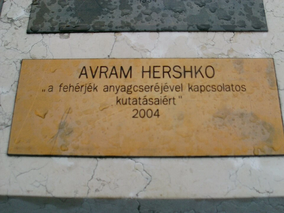
For this historic achievement, Hershko was awarded the 2004 Nobel Prize in Chemistry, making him and Ciechanover the first Israeli citizens to receive a Nobel Prize in the sciences. Later in his career, Hershko expanded his biochemical mastery to elucidate the Anaphase-Promoting Complex / Cyclosome (APC/C), revealing how regulated proteolysis governs chromosome segregation and the eukaryotic cell cycle. This definitive study explores Professor Avram Hershko’s harrowing early life, foundational discoveries, cell cycle breakthroughs, and enduring legacy as one of the world’s greatest experimental biochemists.
1. Childhood in Hungary, Holocaust Survival, and Aliyah to Israel
Avram Hershko (originally Ferenc Hershkó) was born on December 31, 1937, in the small Hungarian town of Karcag, situated in the Northern Great Plain region. His early childhood unfolded against the ominous background of rising European totalitarianism and anti-Jewish legislation enacted in Hungary during the late 1930s. His father, Moshe Hershko, was an esteemed Hebrew teacher at the local Jewish elementary school, and his mother, Shoshana (Margit), was an accomplished pianist. Growing up in a traditional, highly educated Jewish household, young Avram was surrounded by books and scholarly discourse.
Surviving the Holocaust in Austrian Labor Camps
In May 1944, following the Nazi German invasion and occupation of Hungary, the peaceful life of the Karcag Jewish community was shattered. Moshe Hershko was conscripted into the Hungarian forced labor battalions and sent to the frozen Eastern Front in the Soviet Union. Young Avram (then six years old), his mother, and his older brother Chaim were rounded up and forced into the Szolnok ghetto.
While tens of thousands of Hungarian Jews from surrounding regions were deported to the gas chambers of Auschwitz-Birkenau, a fortuitous administrative diversion redirected the train carrying the Hershko family to the Strasshof labor camp in Austria. For nearly a year, Avram and his mother endured severe starvation, freezing temperatures, and forced agricultural labor. Miraculously, both survived the war. Following the liberation of Vienna by Soviet forces in 1945, the family returned to Hungary, where Moshe Hershko miraculously reunited with them after surviving three years of Soviet captivity.
Immigration to Israel and Medical Formations
In 1950, fleeing the stifling constraints of communist totalitarianism and persistent post-war antisemitic sentiment in postwar Hungary, the Hershko family made Aliyah to the newly established State of Israel, establishing their new life in the historic city of Jerusalem. Moshe Hershko resumed his teaching career, authoring foundational Hebrew grammar textbooks, while Avram quickly mastered Hebrew and excelled in the sciences.
In 1956, Hershko enrolled at the Hadassah Medical School of the Hebrew University of Jerusalem. He earned his Master of Science (M.Sc.) in 1965 and his Doctor of Medicine (M.D.) in 1969. During his medical training, Hershko fell under the spell of experimental biochemistry in the laboratory of Professor Jacob Mager, investigating cellular energy metabolism and nucleic acid biosynthesis. Realizing that his true calling lay in basic laboratory discovery rather than bedside clinical practice, Hershko completed his Ph.D. in biochemistry in 1969.
| Academic Stage | Institution | Year | Field / Research Focus |
|---|---|---|---|
| M.D. (Doctor of Medicine) | Hebrew University – Hadassah Medical School | 1969 | Clinical medicine, cellular physiology, pharmacology. |
| Ph.D. Biochemistry | Hebrew University of Jerusalem | 1969 | Enzymatic regulation of cellular nucleotide metabolism under Jacob Mager. |
| Postdoctoral Fellow | University of California, San Francisco (UCSF) | 1969–1971 | Tyrosine aminotransferase (TAT) degradation in cultured hepatoma cells under Gordon Tomkins. |
| Founding Faculty & Chairman | Technion – Rappaport Faculty of Medicine | 1972–Present | Established the Department of Biochemistry; discovered the Ubiquitin System and APC/C. |
2. Postdoctoral Training at UCSF: The Gordon Tomkins Influence
In 1969, Hershko traveled to the University of California, San Francisco (UCSF) for postdoctoral research in the vibrant laboratory of Gordon Tomkins, a charismatic pioneer of molecular endocrinology and enzyme regulation. Tomkins’s lab was investigating the rapid turnover of the enzyme tyrosine aminotransferase (TAT) in rat hepatoma cells.
The Energy Paradox of Proteolysis
While at UCSF, Hershko observed a striking biological paradox: When cultured liver cells were deprived of glucose or treated with mitochondrial inhibitors (like fluoride or dinitrophenol), the rapid degradation of TAT ceased instantly. This was completely counter-intuitive: why should the thermodynamic cleavage of peptide bonds require cellular energy (ATP)?
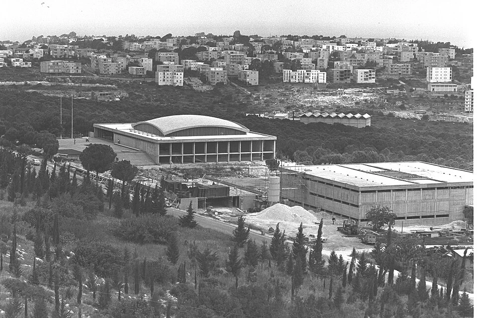
Hershko realized that whole intact cells were too complex to dissect the underlying molecular machinery. To understand the ATP requirement, one had to reconstruct the proteolytic system outside the living cell in a cell-free in vitro system. When Hershko returned to Israel in 1972 to join the fledgling Faculty of Medicine at the Technion in Haifa, he resolved to solve this biochemical mystery.
3. Developing the Reticulocyte Cell-Free System & Fractionation (1972–1978)
At the Technion, Hershko chose an ingenious model system: rabbit reticulocytes (immature red blood cells). As reticulocytes mature into mature erythrocytes, they systematically dismantle and degrade all their internal organelles, ribosomes, and non-hemoglobin proteins. Crucially, reticulocytes have already lost their lysosomes, meaning that any protein degradation occurring within reticulocyte lysate had to be governed by a non-lysosomal, cytosolic machinery.
The DEAE-Cellulose Chromatography Fractionation
In 1977, joined by his brilliant doctoral student Aaron Ciechanover, Hershko subjected the clear reticulocyte lysate to anion-exchange chromatography on a DEAE-cellulose column, separating the lysate into two distinct fractions:
- Fraction I (Unadsorbed): Passed directly through the column without binding to the positively charged resin. It contained hemoglobin and a small, heat-stable polypeptide.
- Fraction II (Adsorbed): Bound tightly to the column resin and was eluted with high salt ($ ext{KCl}$). It contained high-molecular-weight enzymes.
Neither fraction alone could degrade $^{125} ext{I}$-labeled albumin or globin in the presence of ATP. However, when Fraction I and Fraction II were mixed together with ATP and magnesium, robust proteolysis occurred immediately. Hershko named the heat-stable component in Fraction I **APF-1 (ATP-Dependent Proteolysis Factor 1)**.
| Biochemical Component | Original Nomenclature | Molecular Identity | Biological Function |
|---|---|---|---|
| Fraction I Polypeptide | APF-1 (8.5 kDa) | Ubiquitin (76 amino acids) | Covalent molecular destruction tag attached to lysine residues of target proteins. |
| Fraction II Enzyme 1 | Enzyme 1 (E1) | Ubiquitin-Activating Enzyme (UBA1) | Hydrolyzes ATP to activate ubiquitin via adenylation and forms a high-energy E1-thiol ester intermediate. |
| Fraction II Enzyme 2 | Enzyme 2 (E2) | Ubiquitin-Conjugating Enzyme (UBC) | Transfers activated ubiquitin from E1 to its active-site cysteine via trans-thiolation. |
| Fraction II Enzyme 3 | Enzyme 3 (E3) | Ubiquitin-Protein Ligase (UBR / HECT / RING) | Binds specific substrate degrons and catalyzes the isopeptide bond linking ubiquitin to substrate lysines. |
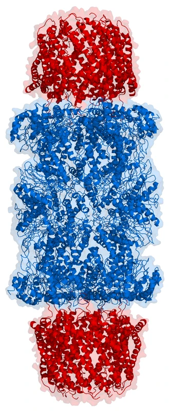
| Megadalton Protease | High-MW Protease Complex | 26S Proteasome (19S + 20S) | Recognizes polyubiquitinated substrates, hydrolyzes ATP to unfold them, and digests them into oligopeptides. |
4. Sabbaticals at Fox Chase and the E1-E2-E3 Enzymatic Cascade
Between 1978 and 1981, Hershko spent summer research sabbaticals at the Fox Chase Cancer Center in Philadelphia in the laboratory of biophysicist and enzymatic mechanism expert Irwin Rose. Rose possessed an extraordinary mastery of isotope exchange kinetics, chemical thermodynamics, and enzyme reaction mechanisms.
The Discovery of Covalent Ubiquitination
Working together at Fox Chase, Hershko, Ciechanover, and Rose made a conceptual leap: APF-1 did not act as an allosteric activator of a protease. Instead, APF-1 was covalently linked to the substrate protein itself.
Through pulse-chase radiolabeling, Hershko demonstrated that multiple APF-1 molecules bound to lysine $epsilon$-amino groups on target proteins via stable isopeptide bonds. The tagged proteins formed high-molecular-weight conjugates that disappeared rapidly as they were hydrolyzed into free amino acids and small peptides, while APF-1 was released intact and recycled. Shortly thereafter, APF-1 was identified as ubiquitin.
Methodical Purification of E1, E2, and E3
Back at the Technion, Hershko undertook the formidable task of purifying the individual enzymatic components of Fraction II using ubiquitin-affinity chromatography (covalently linking ubiquitin to Sepharose beads):
- E1 (Ubiquitin-Activating Enzyme): Hershko purified E1 to homogeneity, demonstrating that it catalyzes a two-step reaction: first forming a ubiquitin-adenylate intermediate ($ ext{Ub}sim ext{AMP}$), then transferring ubiquitin to an internal E1 cysteine to create a high-energy thiol ester bond ($ ext{E1-Cys}sim ext{S-Ub}$).
- E2 (Ubiquitin-Conjugating Enzyme): Hershko isolated multiple distinct E2 isoforms, proving that E2 receives ubiquitin from E1 via trans-thiolation ($ ext{E2-Cys}sim ext{S-Ub}$).
- E3 (Ubiquitin-Protein Ligase): Hershko purified the first E3 ligase (E3$lpha$ / UBR1), demonstrating that E3 is the critical molecular matchmaker: it recognizes specific substrate motifs (such as basic or hydrophobic N-terminal amino acids) and positions the substrate lysine near the E2 active site to form the isopeptide bond.
5. The Biochemical Masterclass: Purifying E1, E2, and E3 to Homogeneity
In the early 1980s, long before the era of recombinant DNA cloning and automated protein purification, purifying low-abundance regulatory enzymes from complex eukaryotic lysates required consummate biochemical skill. Avram Hershko designed an ingenious multi-step purification strategy that remains a classic in enzymatic biochemistry:
1. Covalent Ubiquitin Affinity Chromatography
Hershko chemically conjugated pure bovine ubiquitin to cyanogen bromide (CNBr)-activated Sepharose beads. When crude Fraction II was passed through the column in the presence of $ ext{ATP}$ and $ ext{Mg}^{2+}$, both E1 and E2 enzymes bound tightly to the resin by forming covalent thioester linkages with the immobilized ubiquitin.
Hershko then selectively eluted the enzymes using precise biochemical conditions:
- Elution of E1: Disrupted by high concentrations of AMP and inorganic pyrophosphate ($), which reversed the adenylation reaction, releasing pure 105-kDa E1.
- Elution of E2: Eluted using high concentrations of dithiothreitol (DTT), which cleaved the thioester bonds, liberating multiple distinct E2 species (ranging from 14 to 35 kDa).
- Isolation of E3: Because E3 ligases bind non-covalently to ubiquitin-charged E2 and substrate proteins, Hershko purified the first E3 ligase (lpha$) using sequential gel filtration (Sephacryl S-300) and DEAE-Sephacel chromatography.
2. The Chemistry of Thiol-Ester Intermediates
Hershko demonstrated that the ubiquitin-activating enzyme (E1) catalyzes a remarkable two-step ping-pong mechanism:
- $ ext{E1} + ext{Ub} + ext{ATP}
ightleftharpoons ext{E1}cdot( ext{Ub-AMP}) + ext{PP}_i$ - $ ext{E1}cdot( ext{Ub-AMP}) + ext{E1-Cys-SH}
ightleftharpoons ext{E1-Cys-S}sim ext{Ub} + ext{AMP}$
This was demonstrated through isotopic {32} ext{P} ext{-PP}_i leftrightarrow ext{ATP}$ exchange assays and acid-urea gel electrophoresis, showing that the covalent bond between ubiquitin and E1 was labile to mild alkali and reducing agents, characteristic of a high-energy thioester bond ($sim 34 ext{ kJ/mol}$).
6. Molecular Architecture and Degron Recognition of the APC/C
Following his discovery of the Anaphase-Promoting Complex (APC/C) in 1995, Hershko unraveled the molecular mechanisms by which this giant 1.5-megadalton ligase specifically identifies its mitotic targets while ignoring thousands of other cellular proteins.
The Degron Code: D-Boxes and KEN-Boxes
Hershko demonstrated that substrate proteins targeted by the APC/C possess specific short peptide sequence motifs known as degrons:
- The Destruction Box (D-Box): A conserved 9-amino acid motif with the consensus sequence $ ext{R-x-x-L-x-x-x-x-N}$ (where R is arginine, L is leucine, N is asparagine, and x is any amino acid). The D-box is found in Cyclin A, Cyclin B, and Securin.
- The KEN-Box: A conserved tripeptide motif ($ ext{Lys-Glu-Asn}$ / $ ext{KEN}$) recognized primarily by the $ ext{APC/C}^{ ext{Cdh1}}$ complex in late mitosis and G1 phase, directing the degradation of Nek2, Aurora A kinase, and Cdc20.
- The ABBA Motif & CRY-Box: Secondary degron motifs that fine-tune substrate recruitment affinity during rapid mitotic transitions.
The Multi-Subunit Architecture of the APC/C
Structural biology (Cryo-EM) has confirmed the modular architecture predicted by Hershko’s biochemical dissection:
- The Catalytic Core (Apc2 & Apc11): Apc2 acts as a cullin-like scaffold, while Apc11 is a RING-finger domain that recruits the E2 conjugating enzyme (Ube2C or Ube2S) to catalyze ubiquitin transfer.
- The TPR Scaffold Subcomplex (Apc3, Apc6, Apc7, Apc8): A crescent-shaped array of tetratricopeptide repeat proteins that provides structural rigidity and binds regulatory co-activators.
- The Co-Activator Adaptors (Cdc20 & Cdh1): Possess seven-bladed $ ext{WD40}$ $eta569Xpropeller domains that physically bind the substrate D-box and KEN-box motifs, presenting the substrate directly to the catalytic core.
7. The Spindle Assembly Checkpoint (SAC): Guardian of Mitotic Fidelity
Hershko’s investigations into the APC/C elucidated the biochemical circuitry of the Spindle Assembly Checkpoint (SAC)—the primary surveillance mechanism preventing chromosome mis-segregation during cell division.
If even a single kinetochore on one chromosome fails to attach properly to spindle microtubules from opposite centrosomes, the unattached kinetochore acts as a catalytic factory, generating the Mitotic Checkpoint Complex (MCC) composed of Mad2, BubR1, and Bub3. The MCC binds directly to Cdc20, physically occluding its substrate-binding pocket and sequestering it from the APC/C. This halts anaphase onset with absolute fidelity until tension across all kinetochores is verified, preventing aneuploidy, birth defects, and tumorigenesis.
8. Deciphering the Cell Cycle: The Anaphase-Promoting Complex (APC/C)
Having decoded the basic mechanics of ubiquitin-mediated proteolysis in the 1980s, Avram Hershko turned his attention in the 1990s to an even deeper biological frontier: How does regulated protein degradation control the eukaryotic cell division cycle?
The Mitotic Trigger: How Cells Divide
During mitosis, a mother cell must duplicate its chromosomes and distribute exactly one copy of each chromosome to two daughter cells. If chromosomes separate prematurely or unequally (aneuploidy), the result is embryonic lethality or aggressive cancer. For decades, cell biologists did not know what physical trigger prompted sister chromatids to separate at the transition from metaphase to anaphase.
Discovery and Architecture of the APC/C
In 1995, working with cell-free extracts of clam oocytes and Xenopus frog eggs, Hershko discovered a colossal, 1.5-megadalton multi-subunit E3 ubiquitin ligase: the Anaphase-Promoting Complex / Cyclosome (APC/C) (composed of 14 distinct core subunits).
Hershko demonstrated that the APC/C functions as the master biochemical executioner of mitosis:
- Destruction of Securin: During metaphase, sister chromatids are held together by a molecular glue ring called cohesin. The enzyme that cleaves cohesin—separase—is held inactive by an inhibitor called securin. At anaphase onset, the APC/C polyubiquitinates securin, sending it to the proteasome for destruction. Liberated separase instantly cleaves cohesin, allowing spindle fibers to pull sister chromatids to opposite poles of the dividing cell.
- Destruction of Cyclin B: To exit mitosis and divide into daughter cells, the cell must inactivate its master mitotic kinase, CDK1 (Cyclin-Dependent Kinase 1). The APC/C polyubiquitinates Cyclin B, causing its rapid destruction by the proteasome, which shuts down CDK1 activity and allows the cell to reform its nuclear envelope and undergo cytokinesis.
- Co-activator Regulation (Cdc20 and Cdh1): Hershko decoded how the APC/C changes its substrate specificity during different cell cycle phases by alternating between two regulatory co-activators: Cdc20 in early mitosis and Cdh1 in late mitosis and G1 phase.
| Cell Cycle Event | Substrate Protein | Specific APC/C Complex | Downstream Biological Consequence |
|---|---|---|---|
| Metaphase-to-Anaphase Transition | Securin (Pttg1) | $ ext{APC/C}^{ ext{Cdc20}}$ | Releases separase to cleave cohesin rings; sister chromatids separate synchronously. |
| Mitotic Exit & Cytokinesis | Cyclin B1 / CDK1 complex | $ ext{APC/C}^{ ext{Cdc20}}$ and $ ext{APC/C}^{ ext{Cdh1}}$ | Inactivates CDK1 kinase; permits spindle disassembly, nuclear envelope reformation, and cell cleavage. |
| Spindle Assembly Checkpoint (SAC) | Mitotic Checkpoint Complex (Mad2-BubR1-Bub3) | Inhibits $ ext{APC/C}^{ ext{Cdc20}}$ | Halts anaphase until every single kinetochore is properly attached to bipolar mitotic spindles. |
| G1 Phase Maintenance | Geminin, Plk1, Aurora Kinases | $ ext{APC/C}^{ ext{Cdh1}}$ | Prevents premature DNA re-replication; maintains chromosome integrity prior to S phase entry. |
10. The Structural Superfamilies of E3 Ubiquitin Ligases
Hershko’s original purification of the first E3 ligase (E3-alpha) opened the floodgates to the discovery of more than 600 distinct E3 ubiquitin ligase genes in the human genome (surpassing the ~518 protein kinases). Structural and mechanistic biology has classified these enzymes into three major mechanistic superfamilies:
1. RING (Really Interesting New Gene) and U-box Ligases
The largest class of E3 ligases (comprising over 500 human enzymes, including MDM2, c-Cbl, and the cullin-RING superfamily). RING ligases function as catalytic scaffolds: they bind the ubiquitin-charged E2 enzyme (E2-Ub) and the substrate simultaneously, orienting the substrate lysine residue to attack the E2-Ub thioester bond directly without forming an intermediate covalent bond with the E3 itself.
2. HECT (Homologous to the E6-AP Carboxy Terminus) Ligases
Discovered during studies of human papillomavirus (HPV) E6-mediated degradation of p53, HECT ligases (such as NEDD4, Smurf1/2, and ITCH) possess an active-site cysteine within their conserved 350-amino acid C-terminal domain. Ubiquitin is transferred from E2 to the HECT catalytic cysteine, forming a covalent E3-Ub thioester intermediate before being transferred onto the substrate lysine.
3. RBR (RING-Between-RING) Ligases
A hybrid class (including Parkin and HOIP) containing two RING domains (RING1 and RING2) separated by an in-between-RING (IBR) zinc-binding domain. The RING1 domain recruits E2-Ub, while the RING2 domain carries a catalytic cysteine that forms a covalent thioester intermediate, combining RING recruitment with HECT-like catalysis.
| E3 Ligase Superfamily | Catalytic Mechanism | Representative Human Ligases | Associated Diseases & Pathologies |
|---|---|---|---|
| Cullin-RING Ligases (CRLs) | Scaffold-mediated direct transfer from E2 to substrate. | SCF-Skp2, SCF-betaTrCP, VHL, CRL4-CRBN | Renal cell carcinoma (VHL loss), multiple myeloma, lymphoma. |
| APC/C Complex | Multi-subunit RING-type ligase with interchangeable co-activators. | APC/C-Cdc20, APC/C-Cdh1 | Chromosomal instability, aneuploidy, aggressive solid carcinomas. |
| HECT Domain Ligases | Two-step transfer via obligatory E3-Cys-Ub thioester intermediate. | UBE3A (E6AP), NEDD4-1/2, ITCH | Angelman syndrome (UBE3A mutation), Liddle syndrome (NEDD4 dysregulation), cervical cancer. |
| RBR (RING-Between-RING) | Hybrid mechanism: RING1 binds E2, RING2 forms thioester. | Parkin (PRKN), HOIP, ARIH1 | Autosomal recessive juvenile Parkinson’s disease, chronic autoinflammation. |
11. Therapeutic Exploitation of Mitotic Proteolysis in Oncology
Avram Hershko’s discovery of the APC/C transformed cancer biology by providing a precise roadmap for targeting the mitotic machinery in rapidly dividing malignant cells:
1. Antimitotic Chemotherapies and Spindle Toxins
Classic chemotherapeutic agents such as taxanes (paclitaxel, docetaxel) and vinca alkaloids (vincristine, vinblastine) function by disrupting microtubule dynamics. This prevents kinetochores from establishing tension, permanently triggering the Spindle Assembly Checkpoint (SAC) and blocking APC/C-Cdc20 activation. Cancer cells remain trapped in mitotic metaphase for hours, ultimately succumbing to mitotic catastrophe and apoptotic cell death.
2. Direct Small-Molecule APC/C Inhibitors
Modern oncology research has developed direct pharmacological inhibitors targeting the APC/C:
- ProTAME: A cell-permeable prodrug that disrupts the association of Cdc20 and Cdh1 with the APC/C TPR subcomplex, arresting cancer cells in mitosis.
- Apcin: Binds competitively to the D-box binding pocket of Cdc20, preventing recognition of Cyclin B and Securin.
- Combination Therapy: Synergy between ProTAME and Apcin produces complete mitotic arrest and tumor regression in preclinical models of taxane-resistant breast and ovarian carcinomas.
12. Mentorship, Technion Eminence, and Scientific Philanthropy
Beyond his epochal research, Avram Hershko is revered as an extraordinary mentor who helped build the Technion’s Ruth and Bruce Rappaport Faculty of Medicine from an ambitious regional medical school into an internationally acclaimed scientific powerhouse.
Hershko has mentored generations of Israeli medical students, doctoral fellows, and postdocs, fostering an intellectual culture based on intellectual rigor, self-reliance, and direct empirical observation. He has donated substantial portions of his international prize endowments to establish research fellowships and advanced instrumentation suites for young Israeli scientists.
12. Evolutionary Biology: From Yeast and Plants to Mammalian Systems
The ubiquitin-proteasome pathway decoded by Avram Hershko represents one of the most astonishing examples of evolutionary conservation in all of molecular biology. The 76-amino acid sequence of ubiquitin is identical between human, bovine, and murine species, and differs by only three conservative amino acid substitutions in baker’s yeast (Saccharomyces cerevisiae)—an astonishing degree of structural invariance across more than one billion years of eukaryotic evolution.
The Plant Ubiquitinome: Hormonal Orchestration
In the plant kingdom, the ubiquitin system expanded to unprecedented dimensions. In the model plant Arabidopsis thaliana, more than 6% of the total genome (over 1,400 genes) encodes components of the ubiquitin system, predominantly F-box proteins within SCF ligase complexes:
- Auxin Signaling: The plant hormone auxin binds the F-box receptor TIR1, acting as a molecular glue that recruits Aux/IAA transcriptional repressors for rapid degradation, derepressing genes that govern plant growth and root patterning.
- Jasmonate Defense Signaling: The defense hormone jasmonate binds the COI1 F-box receptor, triggering the degradation of JAZ repressors to activate botanical defenses against herbivorous insects and necrotrophic pathogens.
Prokaryotic Pupylation: Convergent Evolution
While standard bacteria rely on energy-dependent proteases (ClpXP, Lon, FtsH) without ubiquitin, actinobacteria such as Mycobacterium tuberculosis evolved an analogous system called Pupylation. Instead of ubiquitin, a 64-amino acid Prokaryotic Ubiquitin-like Protein (Pup) is attached to substrate lysines by the ligase PafA, targeting doomed bacterial proteins to the bacterial 20S proteasome (Mpa-proteasome complex)—a key vulnerability currently targeted by novel anti-tuberculosis antibiotics.
13. The 2004 Nobel Prize in Chemistry & Global Recognition
On October 6, 2004, the Royal Swedish Academy of Sciences awarded the Nobel Prize in Chemistry jointly to Avram Hershko, Aaron Ciechanover, and Irwin Rose “for the discovery of ubiquitin-mediated protein degradation.”
| Major Award / Distinction | Awarding Institution / Body | Year | Citation / Significance |
|---|---|---|---|
| Israel Prize in Biochemistry | State of Israel | 1994 | Highest national honor for pioneering discovery of non-lysosomal protein degradation. |
| Gairdner Foundation International Award | Gairdner Foundation, Canada | 1999 | Deciphering the ubiquitin-mediated proteolytic pathway. |
| Albert Lasker Basic Medical Research Award | Albert and Mary Lasker Foundation, USA | 2000 | “America’s Nobel”: for identifying the ubiquitin system of regulated protein degradation. |
| Wolf Prize in Medicine | Wolf Foundation, Israel | 2001 | “For the discovery of the ubiquitin system of intracellular protein degradation.” |
| Nobel Prize in Chemistry | Royal Swedish Academy of Sciences | 2004 | Joint laureate “for the discovery of ubiquitin-mediated protein degradation.” |
14. The Bench-Science Ethos: A Master at the Laboratory Bench
Unlike many senior scientific leaders who transition entirely to administrative, fundraising, or political duties after winning major awards, Avram Hershko is legendary throughout the global scientific community for his unwavering commitment to hands-on bench science.
Well into his seventies and eighties, Professor Hershko could be found every morning in his laboratory at the Technion, wearing a white lab coat, personally pipetting reagents, pouring chromatography columns, fractionating cell lysates, and analyzing radioactive autoradiograms with his own hands. When asked why he continues to perform his own experiments, Hershko famously replied:
“Doing bench science is like playing a musical instrument or painting. If you don’t do it yourself, you lose the feel of nature. The most exciting moments in science happen when an unexpected result appears before your eyes at the bench. If someone else does the experiment, you miss the moment of discovery.”
15. Frequently Asked Questions (FAQ)
What was Avram Hershko’s primary scientific discovery?
Avram Hershko discovered the ubiquitin-proteasome system of targeted intracellular protein degradation and purified the key enzymes (E1, E2, and E3) responsible for attaching ubiquitin tags to target proteins, winning the 2004 Nobel Prize in Chemistry.
How did Hershko’s childhood in the Holocaust shape his life?
Born in Hungary, Hershko survived the Strasshof labor camp in Austria as a young child with his mother. The resilience and discipline forged during these early hardships contributed to his extraordinary patience, focus, and perseverance as an experimental biochemist.
What is the Anaphase-Promoting Complex (APC/C) discovered by Hershko?
The APC/C is a multi-subunit E3 ubiquitin ligase discovered by Hershko in 1995 that controls eukaryotic cell division by targeting securin and Cyclin B for destruction, triggering chromosome separation and mitotic exit.
Why was the reticulocyte cell-free system so important?
Rabbit reticulocytes (immature red blood cells) lack lysosomes but actively degrade non-hemoglobin proteins. Hershko used reticulocyte lysates to establish the first cell-free in vitro system that allowed fractionating the cytosolic enzymes of ubiquitin-dependent proteolysis.
What is Avram Hershko’s philosophy on experimental science?
Hershko champions direct, hands-on bench science, believing that senior researchers must perform their own experiments to maintain an intuitive grasp of biological phenomena and catch serendipitous discoveries that automated assays miss.