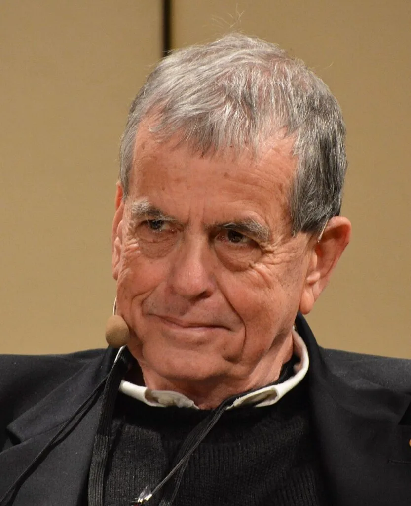
Until the late 1970s, the scientific world viewed the cell through a fundamentally one-sided lens: molecular biologists poured immense resources into unraveling how cells synthesize proteins, transcribe RNA, and duplicate DNA, while virtually ignoring how cells dismantle, regulate, and dispose of their molecular components. Cellular protein degradation was assumed to be a non-specific, passive “garbage disposal” process occurring randomly within acidic lysosomal organelles.
Israeli physician-biochemist Aaron Ciechanover, working alongside his mentor Avram Hershko at the Technion in Haifa and collaborator Irwin Rose at the Fox Chase Cancer Center in Philadelphia, shattered this simplistic assumption. Together, they discovered the Ubiquitin-Proteasome System (UPS)—a highly sophisticated, ATP-dependent molecular tagging and destruction mechanism that functions as the cell’s ultimate quality-control network and regulatory switchboard.
For this historic breakthrough, Ciechanover, Hershko, and Rose were awarded the 2004 Nobel Prize in Chemistry, making Ciechanover and Hershko the first Israeli citizens to receive a Nobel Prize in the sciences. Today, ubiquitin biology forms the cornerstone of modern cancer therapeutics, neurodegenerative disease research, personalized oncology, and next-generation drug modalities such as PROTACs. This comprehensive study explores Professor Aaron Ciechanover’s life, scientific discoveries, clinical impacts, and philosophical leadership in biomedical ethics.
1. Early Life, Family Roots, and Medical Education
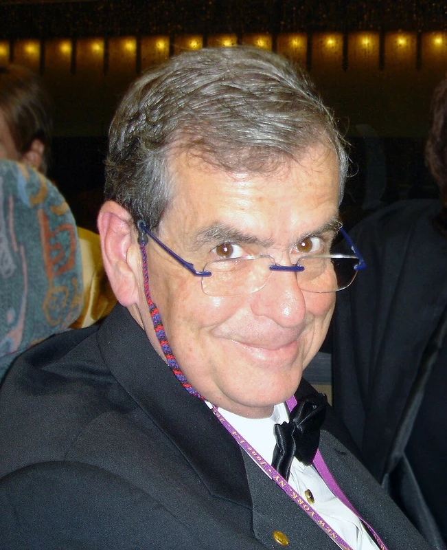
Aaron Ciechanover was born on October 1, 1947, in Haifa, Mandatory Palestine, just seven months prior to the formal declaration of the modern State of Israel. His upbringing coincided with the turbulent, pioneering era of Israeli state-building, characterized by economic austerity, immigrant absorption, and relentless national defense. His parents, Yitzhak and Bluma Ciechanover, were Polish-Jewish immigrants from Ciechanów and Warsaw who had immigrated to Mandatory Palestine in the 1920s and 1930s. His father was an attorney and legal scholar, while his mother was an English teacher and homemaker. Growing up in the picturesque coastal city of Haifa on the slopes of Mount Carmel, Aaron and his older brother Joseph were raised with a profound reverence for Jewish scholarship, literature, and intellectual discipline.
Tragically, Aaron lost both of his parents at a young age: his mother died when he was only ten years old, and his father passed away when he was fifteen. Raised by his older brother Joseph and aunt, Ciechanover channeled his grief into academic excellence. Captivated by biology and human pathology, he collected wildflowers, pressed them into scientific botanical albums, and read avidly about human physiology.
From Medical Doctor to Research Biochemist
In 1965, Ciechanover was accepted into the prestigious academic military program (Atuda) to study medicine at the Hadassah Medical School of the Hebrew University of Jerusalem. He earned his Bachelor of Science (B.Sc.) in 1970 and his Doctor of Medicine (M.D.) in 1972.
Between 1973 and 1976, Ciechanover served as a combat physician and medical officer in the Israel Defense Forces (IDF), including frontline service during the intense combat of the 1973 Yom Kippur War. The harrowing clinical realities of war cemented his realization: while clinical medicine treated the outward symptoms of disease, true medical revolutions could only emerge from understanding disease mechanisms at the fundamental molecular and biochemical level. In 1976, upon completing his military service, Dr. Ciechanover joined the laboratory of Professor Avram Hershko at the newly established Faculty of Medicine at the Technion in Haifa to pursue his doctoral degree (D.Sc.) in biochemistry.
| Academic Degree / Milestone | Institution | Year | Field / Research Focus |
|---|---|---|---|
| B.Sc. & M.D. (Doctor of Medicine) | Hebrew University – Hadassah Medical School | 1970–1972 | Human anatomy, internal medicine, surgery, clinical pharmacology. |
| Military Medical Officer | Israel Defense Forces (IDF) | 1973–1976 | Frontline trauma care, battlefield medicine (Yom Kippur War). |
| D.Sc. Biochemistry | Technion – Israel Institute of Technology | 1981 | Discovery of ATP-dependent ubiquitin-mediated proteolysis under Avram Hershko. |
| Postdoctoral Research Fellow | Massachusetts Institute of Technology (MIT) | 1981–1984 | Cellular biology of transferrin receptor internalization and cell cycle mutants under Harvey Lodish. |
| Professor & Research Director | Technion – Rappaport Faculty of Medicine | 1984–Present | Distinguished Research Professor; Director of the Rappaport Family Institute for Research in Medical Sciences. |
2. The Biological Dogma: Why Protein Destruction Was Overlooked
Throughout the 1950s and 1960s, molecular biology underwent a golden age of structural and biosynthetic discovery: James Watson and Francis Crick elucidated the DNA double helix (1953), the triplet genetic code was deciphered, and protein translation mechanisms were mapped. In this intellectual climate, synthesis was viewed as the sole regulatory mechanism of life.
Biochemists viewed protein breakdown as a purely passive, thermodynamically downhill reaction (exergonic hydrolysis of peptide bonds). Christian de Duve’s discovery of the lysosome in 1955 (which earned him the 1974 Nobel Prize) reinforced this paradigm: scientists believed that damaged or aged proteins were engulfed non-specifically into acidic lysosomes containing acid hydrolases (cathepsins) and digested indiscriminately.
The Biochemical Paradox: Why Does Protein Degradation Require ATP?
However, an acute biochemical paradox baffled perceptive researchers: If the cleavage of a peptide bond releases free energy ($Delta G < 0$), why did cellular experiments show that intracellular protein degradation ceased completely whenever metabolic poisons (such as dinitrophenol or cyanide) depleted cellular adenosine triphosphate (ATP)?
Why would a cell expend precious metabolic energy (ATP) simply to destroy proteins that could spontaneously hydrolyze without energy input? Avram Hershko and Aaron Ciechanover realized that ATP was not required for the thermodynamic cleavage of the peptide bond itself, but for an exquisitely controlled, energy-consuming recognition mechanism that selectively tagged specific proteins for destruction while sparing the rest of the cellular proteome.
3. The Discovery of the Ubiquitin Tag (1978–1981)
In 1977, working at the Technion in Haifa with cell-free reticulocyte (immature red blood cell) lysates, Hershko and Ciechanover separated the lysate into two inactive biochemical fractions using ion-exchange chromatography (DEAE-cellulose):
- Fraction I (Unadsorbed): Contained a heat-stable, small polypeptide of unknown function.
- Fraction II (Adsorbed): Contained high-molecular-weight enzymes.
Neither fraction alone could degrade proteins in the presence of ATP. However, when Fraction I and Fraction II were recombined in the presence of ATP and magnesium ($ ext{Mg}^{2+}$), rapid, selective proteolysis was completely restored. Hershko and Ciechanover named the active peptide in Fraction I APF-1 (ATP-Dependent Proteolysis Factor 1).
APF-1 Is Ubiquitin
During summer research sabbaticals in 1978 and 1979 at the Fox Chase Cancer Center in Philadelphia with biophysicist Irwin Rose, Ciechanover radiolabeled APF-1 with Iodine-125 ($^{125} ext{I}$) and monitored its interactions with substrate proteins. The results were revolutionary:
Under ATP hydrolysis, multiple molecules of APF-1 covalently attached to lysine residues on target proteins, forming high-molecular-weight multi-branched ladders. Shortly thereafter, APF-1 was identified as ubiquitin—a highly conserved 76-amino acid polypeptide previously isolated by Gideon Goldstein in 1975, whose exact sequence is virtually identical from yeast to humans (differing by only 3 amino acids out of 76 across a billion years of evolution).
| Enzyme Family | Specific Name & Function | Human Genes | Biochemical Mechanism of Action |
|---|---|---|---|
| E1 Enzyme | Ubiquitin-Activating Enzyme | 2 Genes (UBA1, UBA6) | Hydrolyzes ATP to form a high-energy ubiquitin-adenylate intermediate, then transfers ubiquitin to its active-site cysteine via a high-energy thioester bond. |
| E2 Enzyme | Ubiquitin-Conjugating Enzyme | ~40 Genes (UBE2 family) | Accepts ubiquitin from E1 through a trans-thiolation reaction, shuttling activated ubiquitin to specific E3 ligases. |
| E3 Enzyme | Ubiquitin Ligase (Substrate Recognition) | >600 Genes (RING, HECT, RBR) | Provides substrate specificity. Binds the specific target protein’s degron motif and catalyzes an isopeptide bond between ubiquitin’s C-terminus (Gly76) and a lysine $epsilon$-amino group on the substrate. |
| 26S Proteasome | Proteolytic Degradation Chamber | Multi-subunit complex (~66 proteins) | Recognizes polyubiquitin chains (Lys48-linked), cleaves and recycles ubiquitin via deubiquitinases (DUBs), unfolds the substrate via ATPases, and hydrolyzes it into short peptides (3–25 amino acids) within its catalytic 20S core. |
4. The Ubiquitin-Proteasome Cascade: How the Cell Destroys Proteins
The ubiquitination cascade decoded by Ciechanover and Hershko operates as a hierarchical, three-tiered enzymatic pyramid that couples chemical activation with exquisite substrate specificity:
The Three-Tier Enzymatic Cascade
- Ubiquitin Activation (E1): The E1 enzyme catalyzes the ATP-dependent adenylation of the C-terminal glycine (Gly76) of ubiquitin, generating pyrophosphate ($PP_i$) and AMP. E1 then forms a high-energy thioester bond ($ ext{E1-Cys}sim ext{S-Ub}$).
- Ubiquitin Conjugation (E2): The activated ubiquitin is transferred from the E1 cysteine to the catalytic cysteine of an E2 ubiquitin-conjugating enzyme via a trans-thiolation reaction.
- Substrate Ligation (E3): The E3 ubiquitin ligase recognizes a specific recognition motif (degron) on the target protein. E3 facilitates or directly catalyzes the transfer of ubiquitin from E2 onto the $epsilon$-amino group of a specific lysine residue on the substrate, creating a stable isopeptide bond.
Polyubiquitin Chain Topologies and Cellular Signals
A single ubiquitin tag (monoubiquitination) can serve non-degradative signaling roles (such as endocytosis, histone regulation, or DNA repair). However, for proteasomal degradation, ubiquitin itself is repeatedly ubiquitinated on specific internal lysine residues (forming polyubiquitin chains):
- Lysine-48 (K48) Linkages: The canonical degradation signal. A chain of at least four K48-linked ubiquitins targets the substrate to the 26S proteasome for immediate destruction.
- Lysine-63 (K63) Linkages: Non-degradative signaling governing NF-$kappa ext{B}$ kinase activation, DNA double-strand break repair, and endosomal protein trafficking.
- Lysine-11 (K11) Linkages: Controls cell cycle progression and mitotic exit via the Anaphase-Promoting Complex/Cyclosome (APC/C).
5. The Molecular Architecture of the 26S Proteasome
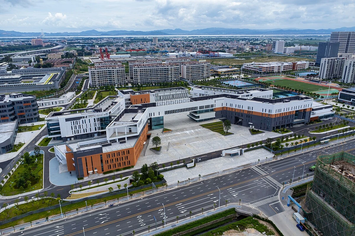
The cellular degradation machine identified by Ciechanover, Hershko, and Rose is the 26S Proteasome—a massive, 2.5-megadalton multi-subunit proteolytic nanomachine composed of approximately 66 individual protein subunits organized into two functional sub-complexes:
1. The 19S Regulatory Particle (The Gatekeeper)
Positioned at one or both ends of the central catalytic cylinder, the 19S regulatory complex performs four indispensable mechanical tasks:
- Ubiquitin Recognition: Specialized ubiquitin receptor subunits (Rpn10, Rpn13, and Rpn1) capture K48-linked polyubiquitin chains on tagged substrates with nanomolar affinity.
- Ubiquitin Recycling (Deubiquitination): The metalloprotease subunit Rpn11 (and associated DUBs like USP14 and UCH37) enzymatically snips the polyubiquitin chain intact from the substrate, recycling free ubiquitin molecules back into the cellular pool.
- ATP-Dependent Protein Unfolding: A hexameric ring of AAA+ ATPases (Rpt1–Rpt6) hydrolyzes ATP to exert mechanical pulling forces, forcefully unfolding the globular, tertiary structure of the target protein into a linear polypeptide string.
- Gate Opening: The C-terminal tails of the Rpt subunits insert into inter-subunit pockets of the 20S core, prying open the narrow central gate to permit entry of the unfolded polypeptide.
2. The 20S Catalytic Core Particle (The Destruction Chamber)
The 20S core is a hollow, barrel-shaped cylindrical structure formed by four stacked heptameric rings (lpha ext{-}7eta ext{-}7eta ext{-}7lpha$):
- Outer $lpha569XRings ($lpha_1–lpha_7$): Form a tightly closed, selective gate preventing non-specific entry of random cytosolic proteins.
- Inner $eta569XRings ($eta_1–eta_7$): Contain the proteolytic active sites pointing inward toward the sealed internal chamber. The active sites employ catalytic N-terminal threonine ($ ext{Thr1}$) residues:
- $eta_1$ Subunit: Caspase-like (peptidyl-glutamyl peptide-hydrolyzing) activity, cleaving after acidic amino acids.
- $eta_2$ Subunit: Trypsin-like activity, cleaving after basic amino acids (Arg, Lys).
- $eta_5$ Subunit: Chymotrypsin-like activity, cleaving after hydrophobic amino acids (Leu, Phe, Tyr). This subunit is the primary therapeutic target of clinical proteasome inhibitors.
6. The N-End Rule Pathway: Decoding Protein Half-Life
In the mid-1980s, Aaron Ciechanover collaborated closely with biochemist Alexander Varshavsky at MIT to resolve another profound biological mystery: What determines why some intracellular proteins persist for days, while others are degraded in minutes?
Together, they discovered the N-End Rule Pathway, proving that the identity of a protein’s amino-terminal (N-terminal) amino acid residue directly dictates its metabolic in vivo half-life:
- Stabilizing Residues (Half-life > 20–30 hours): Methionine, Alanine, Serine, Threonine, Glycine, Valine, Proline.
- Destabilizing Residues (Half-life < 2–30 minutes): Arginine, Lysine, Histidine (basic), Phenylalanine, Tryptophan, Tyrosine, Leucine, Isoleucine (bulky hydrophobic).
Specific E3 ubiquitin ligases (known as N-recognins or UBR proteins) physically bind destabilizing N-terminal residues (primary degrons), triggering immediate polyubiquitination. This provided the first universal biochemical explanation for the regulated lifespan of cellular enzymes, transcription factors, and viral proteins.
| Therapeutic Class | Representative Drug / Molecule | Molecular Target | Clinical Indication / Development Stage |
|---|---|---|---|
| First-Generation Proteasome Inhibitor | Bortezomib (Velcade) | 20S Proteasome $eta_5$ subunit (Reversible) | FDA-approved standard of care for Multiple Myeloma and Mantle Cell Lymphoma. |
| Second-Generation Proteasome Inhibitor | Carfilzomib (Kyprolis) | 20S Proteasome $eta_5$ subunit (Irreversible epoxyketone) | Relapsed / refractory Multiple Myeloma; significantly lower neurotoxicity than bortezomib. |
| Oral Proteasome Inhibitor | Ixazomib (Ninlaro) | 20S Proteasome $eta_5$ subunit (Oral boronate) | First once-weekly oral proteasome inhibitor for combination myeloma therapy. |
| Molecular Glue Degrader (IMiDs) | Lenalidomide / Pomalidomide | Cereblon (CRBN) E3 ligase / IKZF1 & IKZF3 | Reprograms CRBN E3 ligase to degrade oncogenic transcription factors Ikaros/Aiolos; Myeloma & MDS. |
| Clinical PROTAC Candidate | Bavdegalutamide (ARV-110) | Androgen Receptor (AR) / CRBN E3 Ligase | Metastatic castration-resistant prostate cancer (mCRPC) targeting resistant AR point mutants (Phase II). |
| DUB Inhibitor | XL188 / FT671 | USP7 (Ubiquitin Specific Peptidase 7) | Blocks MDM2 stabilization, reactivating endogenous wild-type p53 tumor suppression in solid tumors. |
8. The Expanded Ubiquitin Code: Ubiquitin-Like Modifiers (UBLs) and Non-Proteasomal Signaling
In the decades following the initial discovery, Aaron Ciechanover and international researchers uncovered that ubiquitin is merely the founding member of a vast superfamily of Ubiquitin-Like Modifiers (UBLs) that regulate cellular life without causing protein degradation:
Key Ubiquitin-Like Proteins (UBLs):
- SUMO (Small Ubiquitin-like Modifier): Covalently attached to lysine residues by a specialized E1-E2-E3 cascade. SUMOylation regulates nuclear-cytoplasmic transport, transcriptional repression, genomic stability, and chromosome segregation during mitosis.
- NEDD8 (Neural Precursor Cell Expressed 8): Conjugated to cullin scaffold proteins (NEDDylation) within Cullin-RING E3 ligases (CRLs). NEDDylation induces conformational changes that activate CRL ubiquitination activity, governing over 20% of all cellular proteolysis. The pharmacological NEDD8 inhibitor pevonedistat (MLN4924) is in clinical trials for acute myeloid leukemia.
- ISG15 (Interferon-Stimulated Gene 15): Rapidly induced by Type I interferons during viral infections. ISGylation conjugates to viral and host proteins, directly disrupting viral replication, capsid assembly, and budding in influenza, HIV, and coronaviruses.
- Autophagy Modifiers (LC3 / GABARAP): Cleaved and conjugated to phosphatidylethanolamine (PE) lipids on autophagosomal isolation membranes, driving selective macroautophagy of damaged organelles and protein aggregates.
Linear Ubiquitin Chains and Innate Immunity
Beyond standard isopeptide bonds between the C-terminus of ubiquitin and internal lysine residues, the LUBAC complex (Linear Ubiquitin Chain Assembly Complex) synthesizes head-to-tail linear (M1-linked) ubiquitin chains. These linear chains form specialized scaffold platforms that recruit IKK and NEMO complexes, serving as an essential molecular trigger for innate immune signaling and cell survival during bacterial and viral pathogen exposure.
9. The Technion Rappaport Institute and Israel’s Biomedical Innovation Hub
Aaron Ciechanover’s scientific stature transformed the Ruth and Bruce Rappaport Faculty of Medicine at the Technion into a premier international destination for biochemical and translational medicine. As founding Director of the Rappaport Family Institute for Research in Medical Sciences, Ciechanover established state-of-the-art research facilities spanning cryo-electron microscopy, high-throughput proteomics, and transgenic mammalian modeling.
His academic leadership fostered a culture of translational entrepreneurship, directly stimulating the growth of Haifa’s Matam High-Tech and Life Sciences Park. Technion biomedical graduates and faculty have founded dozens of biotechnology startups pioneering targeted therapeutics, artificial intelligence-driven drug design, and genomic diagnostic platforms, cementing Israel’s standing as a world leader in medical innovation.
10. Vital Physiological Processes Controlled by the UPS
Prior to Ciechanover’s work, protein degradation was thought to be a simple maintenance operation to clear damaged debris. Ciechanover demonstrated that the ubiquitin system is in fact a master regulatory network controlling the precise timing and amplitude of almost every cellular function:
1. Cell Cycle Progression and Mitosis
Orderly cell division requires that regulatory proteins (cyclins and cyclin-dependent kinase inhibitors) appear and disappear at exact microsecond windows. E3 ligases, such as the SCF complex ($Skp1 ext{-}Cullin ext{-}F ext{-box}$) and the APC/C, trigger the rapid destruction of Cyclin A, Cyclin B, and Securin, allowing sister chromatids to separate and enabling the cell to exit mitosis.
2. The p53 Guardian of the Genome Pathway
The master tumor suppressor protein p53 regulates DNA repair, cell cycle arrest, and apoptosis. In healthy cells, p53 levels are kept strictly suppressed by the E3 ubiquitin ligase MDM2, which continuously polyubiquitinates p53 for proteasomal destruction. Upon oncogenic stress or DNA damage, p53 is phosphorylated, disrupting MDM2 binding; p53 rapidly accumulates, halting cell division or triggering apoptosis to prevent malignancy.
3. NF-$kappa ext{B}$ Immune and Inflammatory Activation
The transcription factor NF-$kappa ext{B}$ controls the expression of inflammatory cytokines, chemokines, and cell survival genes. In resting cells, NF-$kappa ext{B}$ is held hostage in the cytoplasm by the inhibitor protein $ ext{I}kappa ext{B}lpha$. Upon inflammatory stimulation (e.g., TNF-$lpha$ or bacterial LPS), the $ ext{I}kappa ext{B}$ kinase (IKK) phosphorylates $ ext{I}kappa ext{B}lpha$, marking it for rapid ubiquitination by $ ext{SCF}^{eta ext{TrCP}}$ and degradation by the proteasome. Liberated NF-$kappa ext{B}$ translocates into the nucleus to activate the immune response.
4. Antigen Presentation & Adaptive Immunity
The 26S proteasome (and its specialized immunological variant, the immunoproteasome) hydrolyzes viral and mutated intracellular proteins into exact 8–11 amino acid oligopeptides. These peptide fragments are transported by TAP into the endoplasmic reticulum, loaded onto Major Histocompatibility Complex (MHC) Class I molecules, and displayed on the cell surface for surveillance by cytotoxic $ ext{CD8}^+$ T lymphocytes.
| Physiological Process | Key Regulatory Protein(s) | Responsible E3 Ligase | Clinical / Biological Outcome |
|---|---|---|---|
| Tumor Suppression | p53 Transcription Factor | MDM2 / HDM2 | Prevents cancer development by triggering apoptosis or senescence upon DNA damage. |
| Mitotic Transition | Cyclin B1, Securin (Pds1) | APC/C (Anaphase Promoting Complex) | Enables separase activation, sister chromatid disjunction, and orderly mitotic exit. |
| Inflammatory Signaling | $ ext{I}kappa ext{B}lpha$ Inhibitory Protein | $ ext{SCF}^{eta ext{TrCP}}$ | Releases active NF-$kappa ext{B}$ to transcribe pro-survival cytokines and immune receptors. |
| Hypoxia Response | HIF-1$lpha$ (Hypoxia Inducible Factor) | VHL (von Hippel-Lindau) | Suppresses angiogenesis under normoxia; allows erythropoietin and VEGF transcription under hypoxia. |
| Mitochondrial Quality Control | Mitofusins (Mfn1/2), VDAC | Parkin (PRKN) | Clears depolarized mitochondria via mitophagy; mutations cause familial Parkinson’s disease. |
11. The UPS in Neurodegenerative Diseases: Protein Misfolding and Aggregate Clearance
In addition to oncogenesis, malfunction of the ubiquitin-proteasome system is intimately linked to the pathogenesis of age-related neurodegenerative diseases characterized by toxic protein aggregation:
Proteasomal Overload in Neuropathology
- Alzheimer’s Disease: Hyperphosphorylated tau protein and amyloid-beta (eta$) oligomers impair the 26S proteasome’s gate-opening mechanics, preventing clearance of misfolded proteins and accelerating synaptic loss and cognitive decline.
- Parkinson’s Disease: Mutations in the E3 ubiquitin ligase Parkin (PRKN) or the mitochondrial kinase PINK1 abolish selective mitophagy of damaged mitochondria. Concurrently, $lpha569Xsynuclein accumulates into ubiquitinated intraneuronal Lewy bodies.
- Amyotrophic Lateral Sclerosis (ALS) & FTD: Mutations in UBQLN2 (ubiquilin-2) or VCP/p97 impair ubiquitin-dependent proteasomal shuttling, causing cytosolic aggregation of TDP-43 and FUS in motor neurons.
- Huntington’s Disease: Expanded polyglutamine (polyQ) repeats in the huntingtin protein resist proteasomal unfolding, physically clogging the 20S catalytic barrel and causing striatal neuronal apoptosis.
Aaron Ciechanover’s recent research investigates small-molecule proteasome activators and autophagy-enhancers that restore cellular clearance capacity in aging neurons, offering transformative therapeutic avenues for neurodegenerative disorders.
12. The 2004 Nobel Prize in Chemistry
On October 6, 2004, the Royal Swedish Academy of Sciences announced that the Nobel Prize in Chemistry 2004 was awarded jointly to Aaron Ciechanover, Avram Hershko, and Irwin Rose “for the discovery of ubiquitin-mediated protein degradation.”
A Historic Triumph for Israeli Science
The award made history: Ciechanover and Hershko became the first Israeli citizens to win a Nobel Prize in the sciences, propelling Israel into the top tier of global biomedical research nations. At age 57, Ciechanover was among the youngest chemistry laureates in decades.
| Major Scientific Award | Awarding Institution | Year | Citation / Significance |
|---|---|---|---|
| Albert Lasker Basic Medical Research Award | Albert and Mary Lasker Foundation, USA | 2000 | “America’s Nobel”: for discovering the ubiquitin system of regulated protein degradation. |
| Israel Prize in Biology | State of Israel | 2003 | Highest national civilian honor for foundational biological discoveries. |
| Nobel Prize in Chemistry | Royal Swedish Academy of Sciences | 2004 | Co-laureate “for the discovery of ubiquitin-mediated protein degradation.” |
| Foreign Associate | National Academy of Sciences (USA) | 2007 | Elected foreign associate for lifetime leadership in biochemistry. |
| Foreign Member | Russian Academy of Sciences & Pontifical Academy | 2008–2010 | International honorary academy appointments and bioethics advisory roles. |
13. Clinical Translation: From Velcade to Targeted Protein Degraders (PROTACs)
The clinical impact of Ciechanover’s discovery has transformed modern hematology, oncology, and pharmacology, validating the ubiquitin-proteasome system as one of the richest sources of pharmaceutical targets in modern medicine.
The Bortezomib (Velcade) Revolution in Multiple Myeloma
In the early 2000s, pharmaceutical researchers developed bortezomib (Velcade / PS-341), a dipeptidyl boronic acid small molecule that reversibly inhibits the chymotrypsin-like $eta5$ catalytic subunit of the 20S proteasome. Because malignant plasma cells (multiple myeloma) produce vast quantities of toxic misfolded immunoglobulins, inhibiting the proteasome causes catastrophic proteotoxic stress, endoplasmic reticulum collapse, and rapid apoptotic death of cancer cells while sparing normal tissues.
Bortezomib and second-generation proteasome inhibitors (carfilzomib, ixazomib) converted multiple myeloma from a rapidly fatal hematological malignancy into a manageable chronic condition, extending median survival from under two years to more than a decade for hundreds of thousands of patients worldwide.
The PROTAC Revolution: Degrading “Undruggable” Targets
Traditional small-molecule pharmaceuticals function as enzymatic inhibitors, requiring deep hydrophobic pockets on proteins to block their catalytic activity. However, more than **85% of the human proteome** consists of non-enzymatic structural, scaffolding, or transcription factor proteins deemed completely “undruggable.”
Ciechanover’s discovery enabled the emergence of PROTACs (Proteolysis Targeting Chimeras). A PROTAC is a bifunctional hybrid molecule consisting of:
- A target-binding ligand that binds any disease-causing protein.
- A flexible chemical linker.
- An E3 ligase-recruiting ligand (such as thalidomide derivatives binding Cereblon, or VH032 binding VHL).
By bringing an endogenous E3 ubiquitin ligase into physical proximity with the target protein, the PROTAC triggers rapid polyubiquitination and complete destruction of the target by the 26S proteasome. Once degradation occurs, the PROTAC molecule dissociates and repeats the cycle catalytically. PROTACs are currently in advanced Phase II/III clinical trials for prostate cancer (ARV-110), breast cancer (ARV-471), and neurodegenerative tauopathies.
14. Personalized Medicine, Genomics, and Bioethical Leadership
In his post-Nobel career, Aaron Ciechanover has emerged as one of the world’s foremost philosophical and scientific advocates for Personalized Genomic Medicine. In his acclaimed international lectures, Ciechanover articulates a future where medicine transitions from empirical “one-size-fits-all” blockbusters to molecularly targeted therapeutics tailored to an individual’s unique genomic and proteomic profile.
Navigating the Ethical Dilemmas of 21st-Century Biotechnology
Ciechanover frequently addresses the complex bioethical challenges arising from high-throughput genome sequencing, gene editing (CRISPR-Cas9), and artificial intelligence in healthcare:
- Genetic Privacy and Discrimination: Preventing employers, insurance companies, and state entities from weaponizing predictive genetic risk data against citizens.
- Equitable Global Access to Precision Medicine: Ensuring that lifesaving, million-dollar gene therapies and personalized biologics are accessible to developing nations and underprivileged populations rather than becoming the exclusive privilege of wealthy elites.
- The Limits of Germline Gene Editing: Differentiating between therapeutic somatic gene therapy to eradicate devastating single-gene genetic diseases (such as sickle cell anemia or Huntington’s chorea) and unregulated eugenic enhancements of human embryos.
- Physician-Patient Relationship in the Algorithmic Age: Emphasizing that while big-data bioinformatics and AI diagnostics provide unmatched predictive power, empathetic human clinical judgment and patient autonomy must remain the ethical foundation of medicine.
15. Frequently Asked Questions (FAQ)
What did Aaron Ciechanover discover that won him the Nobel Prize in Chemistry?
Aaron Ciechanover co-discovered ubiquitin-mediated protein degradation alongside Avram Hershko and Irwin Rose, elucidating the ATP-dependent enzymatic system (E1-E2-E3 cascade) that marks specific proteins with ubiquitin chains for degradation in the 26S proteasome.
Why was ubiquitin-mediated proteolysis such a fundamental discovery?
It proved that protein degradation is not a passive, non-specific lysosomal destruction process, but an essential, energy-consuming regulatory network controlling cell division, tumor suppression (p53), immune signaling (NF-kB), and DNA repair.
How did Ciechanover’s discovery revolutionize cancer treatment?
The discovery enabled the creation of proteasome inhibitors like bortezomib (Velcade), which revolutionized therapy for multiple myeloma and mantle cell lymphoma by inducing toxic proteotoxic collapse in malignant cells.
What are PROTACs and how do they utilize the ubiquitin system?
PROTACs (Proteolysis Targeting Chimeras) are bifunctional molecules that bring disease-causing proteins into physical proximity with endogenous E3 ubiquitin ligases, inducing selective ubiquitination and complete destruction of previously “undruggable” cancer and neurodegenerative proteins.
What is Aaron Ciechanover’s stance on personalized medicine and bioethics?
Ciechanover champions individualized genomic medicine targeting unique patient mutations, while actively lecturing globally on bioethical safeguards regarding genetic privacy, fair global healthcare access, and responsible guidelines for human gene editing.