For more than two decades, the global scientific community considered the three-dimensional atomic mapping of the ribosome—the colossal, asymmetric cellular machine responsible for translating genetic code into the proteins of life—to be an insurmountable biological impossibility. Consisting of hundreds of thousands of tightly packed atoms, dozens of proteins, and long, highly flexible ribosomal RNA (rRNA) chains, the ribosome was deemed too vast, fragile, and dynamic to ever yield high-resolution diffraction crystals.
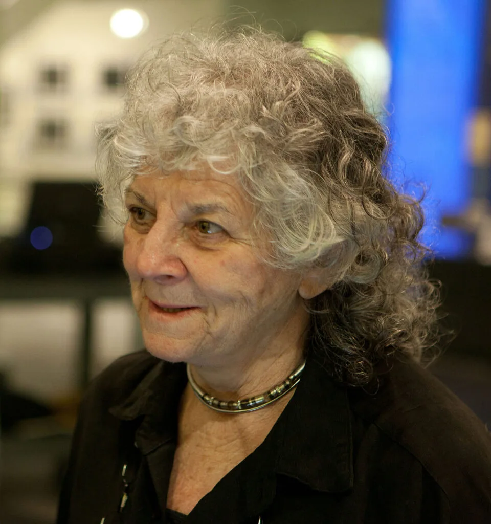
Israeli structural biologist and crystallographer Ada Yonath defied this global scientific consensus. Embarking on what was widely dismissed as a “quixotic fantasy,” Yonath pioneered cryo-bio-crystallography, harvested extremophile bacteria from the Dead Sea and thermal springs, subjected fragile ribosomal crystals to high-energy synchrotron radiation in Hamburg and Grenoble, and ultimately solved the complete three-dimensional atomic architecture of the ribosomal subunits.
Her monumental achievement earned her the 2009 Nobel Prize in Chemistry, making her the first Israeli woman to win a Nobel Prize, the first Middle Eastern woman to win a scientific Nobel, and the first female chemist in 45 years to receive the Nobel distinction since Dorothy Crowfoot Hodgkin in 1964. This definitive study provides an exhaustive examination of Professor Ada Yonath’s life, pioneering methodologies, molecular breakthroughs, antibiotic design paradigms, and her enduring contributions to global biomedical science.
1. Childhood in Jerusalem, Poverty, and Formative Resilience
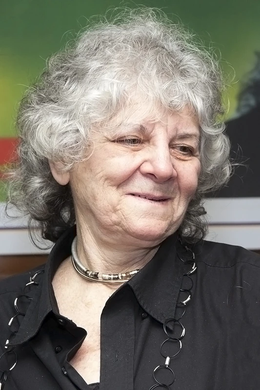
Ada Yonath (née Livshitz) was born on June 22, 1939, in the Geula neighborhood of Jerusalem during the British Mandate. Her parents, Hillel and Esther Livshitz, were Zionist pioneers who had immigrated to Ottoman and Mandate Palestine from Zduńska Wola and Łódź, Poland, in 1933. Her father was an ordained rabbi from a distinguished rabbinical lineage who, unable to secure a rabbinical post, operated a modest neighborhood grocery store.
The family lived in extreme poverty, sharing a cramped four-room apartment with several other families and relatives. Despite constant financial hardship, Yonath’s parents instilled in her an unquenchable thirst for knowledge, literature, and inquiry. When Ada was only five years old, she conducted her first experimental inquiry: attempting to measure the height of the balcony ceiling, she stacked chairs, tables, and crates, climbed to the top, fell, and fractured her arm—a childhood episode she frequently recounts as an early testament to her unstoppable experimental curiosity.
Overcoming Adversity and Scientific Education
Tragedy struck the family when Ada was eleven years old: her father succumbed to severe illness, leaving Esther as a widowed mother with two young daughters in an impoverished Jerusalem. To support her family, young Ada worked ceaselessly—tutoring younger children, cleaning floors, washing dishes, and babysitting—while attending school.
Recognizing her extraordinary intellectual gifts, the prestigious Tichon Beit HaKerem high school in Jerusalem granted her a full scholarship, allowing her to complete her secondary education while continuing to support her mother. Following high school, Yonath completed her mandatory military service in the IDF Medical Corps, where she served in the field hospital unit.
| Academic Stage | Institution | Year | Focus Area / Milestone |
|---|---|---|---|
| B.Sc. Chemistry & Biochemistry | Hebrew University of Jerusalem | 1962 | Organic chemistry, physical chemistry, biochemical pathways. |
| M.Sc. Biophysics | Hebrew University of Jerusalem | 1964 | Macromolecular physical properties, enzyme kinetics, protein crystallization. |
| Ph.D. X-Ray Crystallography | Weizmann Institute of Science | 1968 | High-resolution X-ray structure of collagen under Professor Wolfie Traub. |
| Postdoctoral Research Fellow | Carnegie Mellon University & MIT | 1969–1970 | Muscle protein structural biology (MIT) and macromolecular crystallography (CMU). |
| Founder & Director | Weizmann Institute of Science | 1970–Present | Established Israel’s first biological crystallography laboratory; Director of Kimmelman Center. |
2. The Biological Frontier: What Is the Ribosome?
Every living organism on Earth—from the simplest single-celled bacterium to complex human beings—relies on proteins for virtually every structural, enzymatic, immunological, and signaling function. The blueprint for these proteins is encoded in deoxyribonucleic acid (DNA), transcribed into messenger RNA (mRNA), and delivered to the cellular factory: the ribosome.
The Molecular Complexity of the Protein Factory
Discovered by Romanian-American cell biologist George Palade in the 1950s (for which he won the 1974 Nobel Prize in Medicine), the ribosome is a massive ribonucleoprotein complex comprised of two distinct asymmetrical subunits:
- The Small Subunit (30S in bacteria / 40S in eukaryotes): Responsible for genetic decoding and fidelity. It binds messenger RNA, monitors codon-anticodon pairing with transfer RNA (tRNA), and ensures that genetic instructions are translated without error.
- The Large Subunit (50S in bacteria / 60S in eukaryotes): Responsible for enzymatic catalysis. It contains the Peptidyl Transferase Center (PTC), which catalyzes the formation of peptide bonds between amino acids, synthesizing elongated polypeptide chains that fold into functional proteins.
Unlike simple globular enzymes which consist of a few hundred amino acids, the bacterial ribosome has a combined molecular weight exceeding 2.5 million Daltons, containing more than 50 distinct proteins and three long ribosomal RNA chains totaling over 4,500 nucleotides. Determining its atomic structure required mapping the exact three-dimensional Cartesian coordinates of hundreds of thousands of individual atoms.
3. The Molecular Mechanics of Translation: Initiation, Elongation, Termination
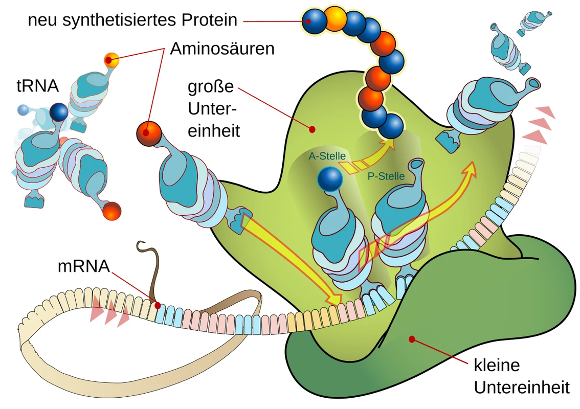
To appreciate the structural marvel decoded by Ada Yonath, one must understand the dynamic four-step cycle of genetic translation executed by the ribosome inside living cells with astonishing speed and accuracy (synthesizing up to 20 peptide bonds per second with error rates lower than 1 in 10,000):
The Three Transfer RNA (tRNA) Binding Sites
The ribosome coordinates three distinct functional binding sites spanning the interface between the small and large subunits:
- The A-Site (Aminoacyl): Binds the incoming aminoacyl-tRNA carrying the specific amino acid corresponding to the mRNA codon being read in the decoding center.
- The P-Site (Peptidyl): Holds the tRNA attached to the growing nascent polypeptide chain. The Peptidyl Transferase Center (PTC) transfers the peptide chain onto the amino acid in the A-site, forming a new peptide bond.
- The E-Site (Exit): Holds the deacylated (uncharged) tRNA after peptide bond synthesis, guiding it out of the ribosome to be recharged by aminoacyl-tRNA synthetase enzymes.
The Translation Cycle Steps
- Initiation: Initiation factors (IF1, IF2, IF3) assemble the 30S subunit, mRNA, and formylmethionine-tRNA (fMet-tRNA) onto the start codon (AUG). The 50S subunit docks to form the intact 70S initiation complex.
- Elongation: Elongation Factor Thermo Unstable (EF-Tu) delivers aminoacyl-tRNA to the A-site. The PTC catalyzes peptide bond formation. Elongation Factor G (EF-G) hydrolyzes GTP to ratchet mRNA and tRNAs forward by exactly one codon triplet (translocation).
- Termination: When a stop codon (UAA, UAG, UGA) enters the A-site, Release Factors (RF1, RF2) bind the decoding center and trigger the PTC to hydrolyze the ester bond, releasing the complete folded protein.
- Recycling: Ribosome Recycling Factor (RRF) and EF-G split the 70S complex into separate 30S and 50S subunits, ready for the next round of protein synthesis.
4. The Quixotic Quest: Overcoming Scientific Scepticism (1980–1999)
In the late 1970s, when Ada Yonath announced her ambition to crystallize the intact ribosome and solve its atomic structure via X-ray crystallography, the global structural biology establishment reacted with overwhelming skepticism. Eminent crystallographers declared the project impossible, labeling Yonath “the village fool” or “a dreamer chasing hallucinations.”
The Fundamental Obstacles
The skepticism was grounded in three formidable physical and biochemical barriers:
- Extreme Conformational Flexibility & Heterogeneity: Ribosomes in living cells exist in continuous dynamic motion, shifting conformations as they bind tRNA, mRNA, and elongation factors. Achieving a population of millions of ribosomes in an identical, rigid conformational state for crystallization seemed unattainable.
- Fragility and Enzymatic Degradation: Ribosomal RNA chains are exceptionally sensitive to minute traces of ubiquitous ribonuclease (RNase) enzymes, causing samples to degrade within hours.
- Catastrophic Radiation Damage: When fragile organic macromolecular crystals are exposed to high-energy synchrotron X-ray beams, the intense ionizing radiation generates free radicals that destroy crystal lattices within seconds, before sufficient diffraction reflections can be recorded.
The Polar Bear Inspiration and Extremophile Strategy
Yonath’s first breakthrough came from an unconventional biological insight: hibernating polar bears. Reading physiological papers on arctic hibernation, Yonath learned that during winter dormancy, polar bear cells systematically pack their ribosomes into tightly ordered, periodically stacked arrays on the inner membranes of their cells, preserving them in a stable, dormant state for months without degradation.
Yonath reasoned that nature had already engineered stable, robust ribosomes capable of withstanding extreme environmental stress. Instead of using fragile laboratory strains of Escherichia coli, Yonath turned to extremophile microorganisms:
- Geobacillus stearothermophilus: A thermophilic bacterium thriving in boiling geothermal hot springs at 65°C.
- Thermus thermophilus: A bacterium discovered in thermal ocean vents.
- Deinococcus radiodurans: An extremophile bacterium isolated from Dead Sea desert soils that survives lethal doses of ionizing radiation, desiccation, and extreme temperatures by tightly protecting its ribosomes.
In 1980, using G. stearothermophilus, Yonath and her team at the Weizmann Institute achieved the world’s first micro-crystals of the large ribosomal subunit (50S)—a historic milestone that silenced early skeptics and initiated a global race.
| Technological Innovation | Pioneered By | Year | Scientific Impact & Legacy |
|---|---|---|---|
| Extremophile Ribosome Isolation | Ada Yonath (Weizmann Institute) | 1980 | Produced thermally stable, structurally homogeneous ribosomal crystals from G. stearothermophilus and T. thermophilus. |
| Cryo-Bio-Crystallography | Ada Yonath & Håkon Hope | 1986–1987 | Flash-freezing crystals to -185°C (88 K) in liquid propane/nitrogen, eliminating radiation decay; now mandatory global standard in structural biology. |
| Synchrotron High-Flux Diffraction | Yonath Group at DESY / EMBL Hamburg & ESRF | 1986–1998 | Harnessed intense synchrotron X-ray beams at particle accelerators, obtaining high-resolution diffraction spots down to 3.0 Ångströms. |
| Heavy-Atom Cluster Phasing | Yonath, Steitz, Ramakrishnan | 1998–2000 | Solved the crystallographic phase problem for millions of atoms using undecagold and tantalum clusters ($ ext{Ta}_6 ext{Cl}_{14}$). |
| Complete 3D Atomic Resolution | Yonath (50S & 30S), Steitz (50S), Ramakrishnan (30S) | 2000–2001 | Published the complete, atomic-coordinate 3D maps of both ribosomal subunits in Cell, Nature, and Science. |
5. The Invention of Cryo-Bio-Crystallography
Even with extremophile crystals, Yonath faced a fatal physical bottleneck: the crystals deteriorated under synchrotron X-ray beams within tenths of a second, destroying diffraction reflections before photographic films or detectors could capture them.
Flash-Cooling at Liquid Nitrogen Temperatures
In the mid-1980s, working in collaboration with Professor Håkon Hope at the University of California, Davis, and utilizing synchrotron beamlines at DESY (Deutsches Elektronen-Synchrotron) in Hamburg, Germany, Yonath developed a revolutionary experimental protocol: Cryo-Bio-Crystallography.
The method involved immersing the delicate ribosomal crystal into a specialized cryo-protectant solution (such as glycerol or methylpentanediol) and plunge-freezing it into liquid propane cooled by liquid nitrogen at **-185°C (88 Kelvin)**. This instantaneous flash-cooling vitrified the surrounding water into amorphous ice without forming crystalline ice shards that would tear the crystal lattice apart.
Global Impact on Structural Biology
At cryogenic temperatures, radiation-induced free radicals are immobilized, extending crystal survival under intense synchrotron beams by more than two orders of magnitude (100-fold). This breakthrough allowed crystallographers to collect thousands of diffraction patterns from a single crystal.
Today, cryo-crystallography is universally employed in every macromolecular crystallography laboratory and synchrotron beamline worldwide, forming the foundational technology behind the resolution of tens of thousands of protein, virus, and enzyme structures in the Protein Data Bank (PDB).
6. The Crystallographic Phase Problem & Heavy Atom Clusters
Recording X-ray diffraction intensities yields only half the information needed to reconstruct an electron density map: it records the amplitude ($|F|$) of scattered X-ray waves, but completely loses the phase angle ($phi$)—a mathematical bottleneck known as the Phase Problem. For a complex as enormous as the ribosome (containing hundreds of thousands of electrons), traditional methods like direct mathematical phasing were completely inapplicable.
Multi-Wavelength Anomalous Dispersion & Giant Heavy Atom Clusters
To overcome the phase problem, Yonath and her contemporaries developed specialized isomorphous replacement and anomalous dispersion protocols using colossal heavy-atom clusters:
- Undecagold Clusters ($ ext{Au}_{11}$): Chemically synthesized multi-gold clusters covalently linked to specific ribosomal protein sulfhydryl groups, serving as massive electron-dense fiducial markers.
- Tantalum Bromide Clusters ($ ext{Ta}_6 ext{Br}_{12}^{2+}$ and $ ext{Ta}_6 ext{Cl}_{14}$): Octahedral heavy metal clusters that diffused into solvent channels between ribosomal subunits without disrupting crystal lattice integrity.
- Synchrotron Wavelength Tuning: By tuning synchrotron X-ray energy precisely to the absorption edges of tantalum, osmium, or selenium atoms, crystallographers recorded anomalous scattering signals, mathematically resolving the phases for over 50,000 diffraction reflections simultaneously.
7. Resolving the 3D Atomic Structure (2000–2001)
Between 1999 and 2001, the intense global race culminated in definitive breakthroughs. Utilizing high-flux synchrotron radiation at DESY in Hamburg and the ESRF (European Synchrotron Radiation Facility) in Grenoble, France, Ada Yonath and her team at the Weizmann Institute achieved high-resolution electron density maps of the large 50S ribosomal subunit from Deinococcus radiodurans at 3.0 Ångströms and the small 30S subunit from Thermus thermophilus.
Simultaneously, independent research groups led by Thomas A. Steitz at Yale University and Venkatraman Ramakrishnan at the MRC Laboratory of Molecular Biology in Cambridge, UK, published high-resolution structures of the 50S subunit from Haloarcula marismortui and 30S subunit from T. thermophilus. Together, these seminal papers revealed the complete mechanical and enzymatic inner workings of the ribosome at single-atom resolution.
Key Architectural Revelations:
- The Ribosome Is a Ribozyme: The structure revealed that the catalytic heart of the ribosome—the Peptidyl Transferase Center (PTC)—contains no protein components whatsoever within 18 Ångströms of the reaction site. The formation of peptide bonds is catalyzed entirely by folded ribosomal RNA (23S rRNA). Proteins serve primarily as structural scaffolds stabilizing the complex RNA core, providing definitive proof for the evolutionary “RNA World” hypothesis.
- The Polypeptide Exit Tunnel: A continuous, non-stick aqueous channel approximately 100 Ångströms long and 15 Ångströms wide traversing the large subunit, through which nascent, newly synthesized protein chains glide outward into the cytoplasm without premature folding or sticking.
- The Decoding Center: Highly conserved adenine nucleotides ($A1492$ and $A1493$ in 16S rRNA) in the small subunit physically flip out to inspect the minor groove geometry of the codon-anticodon pairing, ensuring near-zero error rates during genetic translation.
8. The Chemical Mechanism of Peptide Bond Catalysis in the PTC
One of the greatest debates in 20th-century biochemistry concerned whether the ribosome functioned via chemical acid-base catalysis or purely through structural substrate positioning. Ada Yonath’s high-resolution crystallographic structures, along with subsequent biophysical probing, resolved this question with remarkable clarity.
Entropic Catalysis and Induced Fit
The catalytic strategy employed by the Peptidyl Transferase Center is fundamentally entropic and structural:
- Substrate Proximity and Orientation: The PTC pocket organizes a rigid network of highly conserved ribonucleotides (including $, $, and $) that forms precise hydrogen bonds with the CCA-3′ terminal ends of both A-site and P-site tRNAs. This orients the $lpha569Xamino nucleophile of aminoacyl-tRNA at an optimal 105° Bürgi-Dunitz trajectory toward the carbonyl carbon of the peptidyl ester bond.
- Desolvation and Electrostatic Shielding: By excluding bulk water molecules from the active site cleft, the PTC creates a strictly hydrophobic microenvironment that lowers the activation energy and thermodynamic barrier for zwitterionic and tetrahedral intermediate formation during peptide synthesis.
- Allosteric Signalling and Subunit Coordination: High-resolution crystal structures revealed dynamic inter-subunit bridges (such as Bridge B2a and B3) that transmit structural conformational signals across the 30S decoding center to the 50S catalytic core during tRNA selection.
- Ribose 2′-Hydroxyl Participation: The 2′-OH group of the terminal adenosine (A76) of P-site tRNA acts as an intramolecular proton shuttle, transferring a proton from the attacking amino group to the leaving 3′-oxygen in a concerted six-membered transition state.
| Target Antibiotic | Specific Binding Nucleotide | Primary Resistance Mutation | Biophysical Mechanism of Resistance |
|---|---|---|---|
| Erythromycin / Azithromycin | 23S rRNA adenine 2058 (A2058) | A2058G transition / Erm methylation | Steric clash with desosamine sugar ring; decreases drug affinity by over 10,000-fold. |
| Linezolid | 23S rRNA uridine 2504 & guanine 2576 | G2576U point mutation / Cfr methyltransferase | Disrupts hydrogen bonding network in peptidyl transferase cavity, preventing oxazolidinone docking. |
| Spectinomycin | 16S rRNA helix 34 (cytosine 1092) | C1092U / G1064A mutation | Prevents head rotation locking of 30S subunit required for translocation inhibition. |
| Streptomycin | 16S rRNA 530 loop & protein uS12 (RpsL) | Lys42Arg or Lys87Arg in protein uS12 | Eliminates salt bridge interactions between aminoglycoside and ribosomal decoding pocket. |
9. The Weizmann Institute of Science: An Incubator of Global Structural Biology
Ada Yonath’s landmark achievements cemented the status of the Weizmann Institute of Science in Rehovot, Israel, as an international powerhouse for biomolecular structural research. Established as the Daniel Sieff Research Institute in 1934 by Israel’s first President, Chaim Weizmann (himself an eminent chemist), the institute fosters an academic environment of complete intellectual freedom and state-of-the-art infrastructure.
As Director of the Helen and Milton A. Kimmelman Center for Biomolecular Structure and Assembly, Yonath established a world-class center of excellence that trained generations of Israeli and international structural biologists. The center operates cutting-edge crystallization robotics, high-resolution cryo-electron microscopy (Cryo-EM) suites, and high-field nuclear magnetic resonance (NMR) spectrometers, maintaining strong cooperative ties with international synchrotrons including DESY (Hamburg), ESRF (Grenoble), and the Swiss Light Source (SLS).
10. The 2009 Nobel Prize in Chemistry
On October 7, 2009, the Royal Swedish Academy of Sciences awarded the Nobel Prize in Chemistry jointly to Ada E. Yonath, Venkatraman Ramakrishnan, and Thomas A. Steitz “for studies of the structure and function of the ribosome.”
| Prestigious Award | Awarding Organization | Year | Citation / Achievement |
|---|---|---|---|
| Israel Prize in Chemistry | State of Israel | 2002 | Highest national honor for pioneering structural biology of the protein synthesis machinery. |
| Harvey Prize in Science | Technion – Israel Institute of Technology | 2002 | Breakthrough crystallography of ribosomal particles and ribosome-antibiotic complexes. |
| Wolf Prize in Chemistry | Wolf Foundation, Israel | 2006 | “For ingenious structural discoveries of the ribosomal machinery of peptide-bond formation.” |
| Paul Ehrlich and Ludwig Darmstaedter Prize | Paul Ehrlich Foundation, Germany | 2007 | Deciphering the structure of the ribosome and the mode of action of ribosomal antibiotics. |
| L’Oréal-UNESCO For Women in Science Award | UNESCO & L’Oréal Foundation | 2008 | Laureate for Africa and the Arab States for structural insights into protein synthesis and antibiotics. |
| Nobel Prize in Chemistry | Royal Swedish Academy of Sciences | 2009 | Joint laureate “for studies of the structure and function of the ribosome.” |
11. Transforming Antibiotic Design & Combating Superbug Resistance
More than 50% of all clinically prescribed antibiotics target the bacterial ribosome to arrest pathogenic bacterial growth. Prior to Yonath’s structural breakthroughs, pharmaceutical companies developed antibiotics through empirical trial-and-error chemistry without knowing the exact atomic binding pockets of drug molecules.
Atomic-Resolution Mapping of Clinical Antibiotics
Following the determination of the native ribosomal structure, Yonath crystallized ribosomes complexed with more than 20 major classes of clinically essential antibiotics, uncovering their precise mechanisms of action:
| Antibiotic Class / Drug | Target Subunit & Binding Site | Mechanism of Action | Clinical Applications |
|---|---|---|---|
| Macrolides (Erythromycin, Azithromycin) | 50S Subunit (Upper Exit Tunnel) | Physically plugs the polypeptide exit tunnel, halting protein elongation after 4–8 amino acids. | Respiratory infections, pneumonia, strep throat, skin infections. |
| Aminoglycosides (Gentamicin, Streptomycin) | 30S Subunit (Decoding Center) | Locks decoding adenines in active conformation, causing massive mistranslation of toxic misfolded proteins. | Severe Gram-negative sepsis, hospital-acquired bacterial infections. |
| Tetracyclines (Doxycycline, Minocycline) | 30S Subunit (A-Site) | Sterically blocks aminoacyl-tRNA from entering the A-site, arresting translation initiation. | Lyme disease, acne, cholera, anthrax, multi-drug resistant pathogens. |
| Oxazolidinones (Linezolid) | 50S Subunit (Peptidyl Transferase Center) | Distorts PTC orientation, preventing formation of initiation complex with fMet-tRNA. | Methicillin-resistant Staphylococcus aureus (MRSA), vancomycin-resistant enterococci (VRE). |
| Chloramphenicol | 50S Subunit (PTC A-Site Cleft) | Competitively inhibits peptide bond catalysis by interfering with aminoacyl-tRNA positioning. | Bacterial meningitis, typhoid fever, refractory infections. |
Rational Drug Design against Superbugs
Yonath’s ongoing research at the Weizmann Institute focuses on developing species-specific, pathogen-selective antibiotics. Because human cells contain mitochondrial ribosomes that share structural features with bacterial ancestors, broad-spectrum antibiotics often cause mitochondrial toxicity and destroy beneficial human microbiome flora.
By mapping structural differences in peripheral rRNA loops unique to pathogenic bacteria (such as Mycobacterium tuberculosis, Acinetobacter baumannii, and Pseudomonas aeruginosa), Yonath’s laboratory is engineering next-generation antibiotics that target unique pathogen pockets, preserving the gut microbiome and overcoming mutational antimicrobial resistance.
12. The Proto-Ribosome Hypothesis: The Origin of Life
In analyzing the three-dimensional architecture of the catalytic Peptidyl Transferase Center across bacteria, archaea, and eukaryotes, Ada Yonath discovered a profound structural feature: a universal, semi-symmetrical pocket composed of approximately **180 ribonucleotides** that is identical in sequence and tertiary fold across all forms of life.
The Primordial Molecular Machine
Yonath formulated the “Proto-Ribosome” hypothesis, proposing that before the evolution of genetic code, messenger RNA, or proteins, a small dimeric RNA pocket formed spontaneously in prebiotic oceans. This proto-ribosome possessed the simple catalytic ability to bind two activated amino acids and link them together via peptide bonds, producing random oligopeptides that stabilized early biochemical systems.
Over evolutionary epochs, nature preserved this ancient RNA catalytic engine at the core of the ribosome, gradually adding concentric layers of rRNA and ribosomal proteins to enhance decoding precision and processivity. In her laboratory at the Weizmann Institute, Yonath’s team successfully synthesized artificial mini-RNA constructs mirroring the proto-ribosome, demonstrating that these prebiotic RNA pockets spontaneously catalyze peptide bond synthesis in vitro, providing compelling evidence for how life transitioned from chemical chaos to biological self-replication.
13. Mentorship, Women in STEM, and Scientific Diplomacy
Ada Yonath has emerged as an iconic global role model for women in science, relentlessly encouraging young female students, postdocs, and researchers to pursue careers in chemistry, physics, and biotechnology without compromising their intellectual ambitions or personal lives.
The Philosophy of Scientific Curiosity
Yonath frequently emphasizes that genuine scientific breakthroughs require passion, perseverance, and intellectual independence rather than chasing career metrics or conformity:
“Science is not about money or prestige; it is about curiosity and the joy of unraveling nature’s most profound secrets. When you encounter skepticism or failure, you must believe in your data, stay persistent, and dare to venture into the unknown.”
Through the Weizmann Institute’s educational programs, the Israel Academy of Sciences and Humanities, and international forums such as the Lindau Nobel Laureate Meetings, Yonath mentors hundreds of aspiring scientists worldwide, advocating for equitable funding, family-friendly academic environments, and science education for underprivileged communities.
14. Technological Legacy: From X-Ray Crystallography to Single-Particle Cryo-EM
The cryogenic preservation principles established by Ada Yonath in the 1980s laid the essential groundwork for the modern revolution in Single-Particle Cryo-Electron Microscopy (Cryo-EM), honored with the 2017 Nobel Prize in Chemistry (awarded to Jacques Dubochet, Joachim Frank, and Richard Henderson).
Today, researchers routinely flash-freeze macromolecular complexes in vitreous ice at liquid ethane/nitrogen temperatures without crystallization, capturing heterogeneous functional states of human 80S ribosomes, mitochondrial ribosomes, and spliceosomes at sub-2.0 Ångström resolutions. This technological continuity directly links Yonath’s early cryogenic experiments at the Weizmann Institute to contemporary frontiers in structural biophysics, drug discovery pipelines, and AI-driven macromolecular modeling.
15. Frequently Asked Questions (FAQ)
What did Ada Yonath discover that won her the Nobel Prize in Chemistry?
Ada Yonath decoded the three-dimensional atomic structure and catalytic mechanisms of the ribosome—the cellular factory that synthesizes proteins—using high-resolution X-ray crystallography, sharing the 2009 Nobel Prize in Chemistry with Venkatraman Ramakrishnan and Thomas A. Steitz.
What is cryo-crystallography and why is it so important?
Invented by Ada Yonath in the 1980s, cryo-bio-crystallography involves flash-freezing macromolecular crystals in liquid propane/nitrogen at -185°C to prevent catastrophic radiation damage from high-energy synchrotron X-ray beams. Today, it is the universal standard technique in structural biology laboratories worldwide.
Why was solving the structure of the ribosome considered impossible?
The ribosome is an asymmetric macromolecular complex with a molecular weight of over 2.5 million Daltons, containing hundreds of thousands of atoms, flexible RNA chains, and dozens of proteins. Its conformational instability, vulnerability to enzymatic degradation, and rapid decay under X-rays caused leading scientists to declare it impossible to crystallize.
How has Ada Yonath’s work contributed to modern medicine and antibiotics?
Yonath solved the atomic-level binding structures of over 20 clinical antibiotics targeting the ribosome (including erythromycin, azithromycin, linezolid, and tetracyclines). This knowledge enabled rational drug design to create species-specific antibiotics that fight drug-resistant superbugs while sparing human microbiome flora.
What is the Proto-Ribosome hypothesis proposed by Ada Yonath?
Yonath discovered a symmetrical, universal 180-nucleotide RNA pocket at the core of the peptidyl transferase center present in all living cells. She hypothesized that this ancient RNA entity—the “proto-ribosome”—preceded all cellular life and genetic code, spontaneously linking amino acids in prebiotic oceans.